

Vascular Cryoablation
 Home
Home- Vascular Cryoablation


Medically Reviewed By Dr. Antariksh Vijan Updated on September 08, 2026
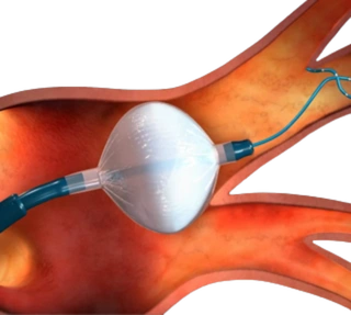
Cryoablation is a minimally invasive treatment that destroys abnormal tissue by freezing it. A trained intervention radiologist places a thin probe or needle in the target area, cools it with cold compressed gas, and the freeze-thaw process kills the cells. The treatment can be used for cancers, pain-related nerve targets or vascular malformations.
In this article, we will be examining cryoablation for vascular malformations. It is emerging as a treatment for low-flow venous malformations and related anomalies, but is still niche and limited to super-speciality centres.
Conditions Treated With Cryoablation
Cryoablation uses controlled freezing to destroy abnormal tissue. It is used across different medical specialties, and the suitability of treatment depends on the type, size and location of the abnormal tissue and whether it can be safely targeted under imaging guidance.
Venous Malformations: Cryoablation is increasingly used for selected symptomatic venous malformations, particularly focal lesions that cause persistent pain, swelling, tenderness or restriction of movement. It may be considered as a primary treatment in carefully selected cases or when treatments such as sclerotherapy have not provided sufficient symptom relief.
Fibroadipose Vascular Anomaly (FAVA): Cryoablation may also be used in selected patients with fibroadipose vascular anomaly (FAVA). FAVA commonly affects muscles and can cause persistent pain, stiffness and limitation of movement. Cryoablation can target a focal painful area while helping preserve the surrounding structures.
Cancerous Tumors: Image guided cryoablation is also used to treat selected cancerous tumors, particularly when the tumor can be precisely targeted without causing unacceptable damage to nearby structures. Depending on the individual case, it may be used for selected tumors involving the kidney, liver, lung, prostate or bone, either as a primary local treatment or alongside other cancer therapies.
Benign Tumors and Painful Lesions: Certain non-cancerous tumors and focal abnormal tissues can also be treated with cryoablation. These include selected benign bone and soft-tissue lesions, particularly when treatment is required to control pain or other symptoms.
Although cryoablation has applications in cancer treatment and other conditions, this article focuses specifically on its use for venous malformations and selected vascular anomalies.
Indications for Cryoablation
As explained above, the procedure is for low-flow venous malformations. It is considered when the symptoms are significant enough to limit daily life — pain or tenderness, a noticeable swelling or lump, and limited movement or function due to the location of the lesion.
Cryoablation is considered when first-line treatments like sclerotherapy do not provide sufficient relief. It’s less likely to be recommended if the lesion is very diffuse, wraps around a nerve or joint. So, anatomy also plays a huge role in selecting treatment. Published outcomes for cryoablation of venous malformations have been encouraging.
Reference: Marcelin C, Linet T, Jambon E, et al. Percutaneous image-guided cryoablation of venous malformation and fibro-adipose vascular anomaly: prognostic factors of clinical efficacy. European Radiology. 2025;35(10):6554–6563. DOI: 10.1007/s00330-025-11545-w.
Steb-by-Step Cryoablation Procedure
- The first step is to check the lump or swelling with an ultrasound, MRI or CT to see its exact size and location. Depending on where it's located, a type of anesthesia is selected — regional or general.
- General anesthesia is relatively common when the lesion is large, deeply located, extremely painful and in a very sensitive place. Also, if the procedure is dependent on the patient remaining still for a longer period of time. If the location is near very important nerves or skin that needs very careful control, general anesthesia is preferred. Local/regional anesthesia is used for less complex lesions that are small, superficial (close to the skin) and in a safe location that can be numbed well with an injection (for e.g., in the leg or arm).
- After anesthesia, vascular & interventional radiologist place one or more cryoprobes — this is a thin needle-like instrument that is used to freeze the abnormal tissue. They use live imaging for guidance. Once the target area has been reached, the probe creates an ‘ice ball’. The tip becomes extremely cold due to cold gas or fluid circulating inside the probe.
- A controlled freeze-thaw cycle is started. The ‘ice ball’ ends up covering the painful part of the malformation and destroys the abnormal tissue.
- An important part of the procedure is to ensure this damage does not spread to surrounding tissues that are normal and functioning. Skin, nerves and other important structures need to be protected from this. To this effect, the operating team watches closely and sometimes uses warm saline or other protective steps.
- Once the procedure is completed, the probe is thawed and removed and the area is observed for numbness, swelling or skin irritation.
- The overall objective is to reduce pain, lump size and overall discomfort and not remove every bit of the malformation — which can be challenging, at times, depending on the placement.
Advanced Techniques in Cryoablation
Advances in cryoablation for vascular purposes revolve around precision. Malformations can be tricky to get to, and live imaging facilitates precision, even before the actual freezing begins. While one probe normally does the job, the use of multiple probes to cover larger malformations is becoming more common. Another major advance is thermocouple monitoring — tiny temperature sensors that are placed near the surrounding nerves to reduce any potential risk. Real-time ice ball monitoring is also used to improve the precision of the treatment. Doctors watch the freeze zone and adjust time and power consistently to avoid harming any healthy tissue.
Benefits of Cryoablation
The primary benefit is reduced pain. Most studies report a dramatic drop in aching, heaviness and pressure or tenderness. There is also a significant reduction in lump size. Most patients show a high degree of improvement, while some experience a complete relief from symptoms.
Another natural benefit is the improved movement and daily function. When the lump is in the hand or leg, or around a specific joint, shrinking it can make certain previously-painful movements possible. Walking, jogging, using a limb, even carrying some load or motor-skill related tasks like tying shoelaces may become easier. Patients show a significant improvement in overall quality of life.
The procedure is minimally invasive, so recovery is always quicker as compared to any invasive procedure. There might be temporary pain, bruising, swelling or some numbness as after-effects of the procedure, but these quickly subside (within a few days to a couple of weeks, at most).
Reference: Fish A, Moushey A, Chan SM, Staib L, Marino A, Schlachter T. Cryoablation of Venous Malformations: A Systematic Review. Journal of Vascular and Interventional Radiology. 2022;33(8):993–1000. DOI: 10.1016/j.jvir.2022.04.010.
Risks and complications of Cryoablation
If the lesions are close to the surface, skin blistering is a concern. Nerve injury from inaccurate freezing is another concern. The surrounding area may feel numb, burning or tingling, feel unusually tender or painful to touch, or if a motor-nerve is involved, some weakness can show. Bleeding or hematoma at the probe site is another risk. Infection is a risk too, but rather uncommon, when proper sterilization technique is employed. If the freezing spreads beyond the target area, there is a risk of damage to nearby tissues. To avoid this, doctors use real-time imaging, saline barriers and temperature monitoring.
There are rarer complications in the case of deep or complex lesions, such as larger skin blisters, nerve symptoms or delayed healing. In one recorded case, a dermal blister (a fluid bubble right under the skin’s outer layers) was caused, but healed on its own within 6 weeks.
All said, it’s one of the safer procedures, with a generally low risk margin.
References:
1. Fish A, Moushey A, Chan SM, Staib L, Marino A, Schlachter T. Cryoablation of Venous Malformations: A Systematic Review. Journal of Vascular and Interventional Radiology. 2022;33(8):993–1000. DOI: 10.1016/j.jvir.2022.04.010. - PubMed: https://pubmed.ncbi.nlm.nih.gov/35469956/
2. Marcelin C, Linet T, Jambon E, et al. Percutaneous image-guided cryoablation of venous malformation and fibro-adipose vascular anomaly: prognostic factors of clinical efficacy. European Radiology. 2025;35(10):6554–6563. DOI: 10.1007/s00330-025-11545-w.
Full text: https://pmc.ncbi.nlm.nih.gov/articles/PMC12456970/
PubMed: https://pubmed.ncbi.nlm.nih.gov/40185927/
Recovery and Rehab
After vascular cryoablation, recovery is fairly quick. Many patients go home on the same day or after a short observation period. However, the exact timeline depends on the complexity and size of lesion that was treated.
In the first 24 hours, it’s important to rest, keep the probe area clean and use pain medication. Some tenderness can be expected during the first week. During this time, vigorous exercise, heavy lifting and longer periods of athletic activity are not recommended. From the second week onwards, patients return to normal life and even light athletic activity, based on the symptoms.
By 4-6 weeks, most patients return to regular activity and exercise routines (higher-impact sports may be avoided for a little longer, but this too is dependent on the individual’s training/case history). However, this is dependent on healing, symptoms and the treatment site.
Rehab is usually relatively simple — light walking, gradual return to activity and follow up scans to check whether the lesions are shrinking. Based on the location of the lesions, doctors may place some limitations on specific movements. For example, if the lesions were near the foot, there might be a limitation on range of movement to expedite healing and reduce swelling.

