

Aortic Dissection
 Home
Home- Aortic Dissection

Medically Reviewed By Dr. Meghav Shah Updated on February 23, 2026
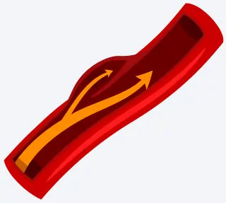
Aortic dissection is a potentially life-threatening condition. A tear develops in the inner-layer of the aorta, allowing blood to flow between the aortic wall layers; this, in turn, can compromise organ perfusion (the process of delivering nutrient-rich blood to the organs and tissues).
When there is a tear, a ‘false channel’ or false lumen. This in turn, interferes with the original channel of blood flow. Over time, as the tear increases, the true lumen can get compressed, in turn starving organs of oxygenated blood.
The Aorta has three layers: intima (inner), media (middle) and adventitia (outer). The tear starts in the intima; high-pressure blood forces its way into the media, and forms a false lumen, parallel to the true one. Eventually, this can rupture outward into the adventitia, driven by shear stress, wall weakness or inflammation.
Causes of Aortic Dissection
Aortic dissection, as explained above, occurs from a tear in the innermost part of the Aorta. However, this does not happen spontaneously. It’s usually a result of a systematic weakening of the aortic wall, in particular the media layer.
This compromise in structural integrity can come from multiple factors like atherosclerosis, Marfan's Syndrome and other forms of trauma.
The aortic wall comprises elastic fibres, smooth muscle cells and extracellular matrix. These undergo degenerative changes, collectively referred to as cystic medial necrosis. This leads to elastin degradation, decreases collagen synthesis and apoptosis (death of cells) of smooth muscle cell layers. These changes give rise to ‘cystic’ spaces occupied by mucoid material, replenished by chondroitin sulfate, thus compromising the elasticity and tensile properties of the aorta, thereby increasing a risk of aortic aneurysm formation and intimal rupture.
Role of Hemodynamic Factors:
In 70-80% of cases, hypertension is an important factor. High blood pressure exerts chronic pulsatile shear stress and wall tension, directly contributing to intimal tear. These tears often occur at high-shear sites, such as the right anterolateral ascending aorta.
Acute spikes, including those from blunt chest trauma and vasoconstriction (cocaine-induced) can precipitate acute failure in a predisposed wall.
Atherosclerosis also has a role, even if that’s lesser than previously thought. It leads to an inflammation and thinning of the medial layer, through rupture of plaques or occlusion of the vasa vasorum (blood vessels supplying the walls of arteries with oxygen and nutrients), which impairs nutrition. Direct injury from trauma, such as in car accidents from deceleration, directly shears the intimal layer.
Sometimes, weakening of blood vessel walls from cardiac catheterization or from inflammatory disease such as Takayasu arteritis (different from arthritis, please note), contribute to the overall deterioration of the blood vessels, making them more susceptible to dissection even under normal pressure.
Risk Factors for Aortic Dissection:
Hypertension is the most common modifiable risk factor. 70-80% of aortic dissections are caused by this. It has also been linked to a higher association with distal dissections (Type B dissections). In Type B dissections, shear stress progressively compromises the media layer. Systolic pressures above 180mmHg, which are sustained, increase tension through the Law of Laplace (Tension = Blood Pressure x Radius/wall thickness). Essentially, this means, a thinner wall will experience significantly more stress at a given pressure, making it prone to tearing. Conversely, high blood pressure affects the stress on the aortic wall, making it prone to the propagation of a dissection.
When the aorta is dilated, the risk of a dissection in hypertensives is doubled.
Genetic predispositions:
The risk is exponentially higher for those with connective tissue disorders, in particular Marfan Syndrome with FBN1 gene mutations. Vascular Ehlers-Danlos syndrome leads to vessel fragility, increasing medium vessel ruptures associated with aortic dissections and tears. Bicuspid aortic valve, present in 0.5-2% of the Indian population (as extrapolated from global data), accelerates abnormal aortic blood flow, significantly increasing the risk in the population with this trait.
Lifestyle factors:
Smoking massively compounds the risk, as is the case with any cardiovascular symptom. Smoking promotes atherosclerosis and MMP activation, which in turn exacerbate the risk of tears. Incidences of aortic dissection peak over the age of 60 (63 years for Type A). Males are 2-3 times more susceptible, perhaps due to testosterone-driven growth and pressure effects.
While not extensively reported, it is important to understand that the use of cocaine massively precipitates Type B dissections in young males due to acute hypertension and catecholamine surges. Drug usage itself is underreported, and screenings tend to avoid looking for it, but this is necessary awareness.
Symptoms of Aortic Dissection
Aortic dissection typically presents with abrupt, excruciating pain that patients describe as ripping, tearing, or stabbing, distinguishing it from the gradually developing pains that characterize myocardial infarction.
Pain Symptoms:
The hallmark of acute type A dissection is extreme pain that reaches a peak at the onset of chest pain. This occurs in 90-96% of patients. Typically anterior (front) chest pain for proximal dissections (Type A) or interscapular/back pain for distal dissections (Type B). The pain may migrate as the dissection propagates; for E.g, chest to abdomen or legs, indicating the extension of the false lumen.
Associated symptoms include nausea, feelings of impending doom, anxiety, diaphoresis, pallor or even episodes of syncope (blackouts) due to transient hypotension (low blood pressure) or vagal behaviour.
Ischemic and Vascular Signs:
Ischemic signs are often characterized by organ-specific deficits. This means, the functioning of various organ systems will change due to vessel occlusion, in 20-30% of cases: unequal pulses (>20mmHg arm discrepancy in 15-30%), limb weakness/ischemia (cold extremities, paresthesia, paresis), stroke symptoms (hemiparesis — muscle weakness/partial paralysis, aphasia — impaired communication ability, vision loss), or abdominal pain related to mesenteric ischemia.
The patient will have organ-specific deficits due to vessel occlusion in 20-30% of cases: unequal pulses/blood pressures (>20mmHg arm discrepancy, 15-30%), limb weakness/ischema (cold extremities, paresis, paresthesia), stroke symptoms (hemiparesis, aphasia, vision loss), or abdominal pain related to mesenteric ischemia.
In 5-10% of cases, myocardial ischemia can mimic STEMI (ST-elevation myocardial ischemia — essentially, a type of severe heart attack) through the involvement of coronary ostia. Dyspnea and orthopnea arise because of pericardial (heart membrane) effusion/tamponade.
Physical Findings:
Physical examination may show pulse deficits (~30%), Type A aortic regurgitant murmur, Horner syndrome if the carotid sinus is involved, or hoarseness if the RL (Recurrent Laryngeal) nerve is involved. Neurological deficits account for about 20%, through paraplegia from spinal cord ischemia. Painless presentations (asymptomatic) are often misdiagnosis risks, getting mistaken for syncope or coma.
Diagnosis of Aortic Dissection
It involves pre-emptive, high suspicion and immediate screening, due to the mimicking nature of the symptoms. They can resemble many different conditions. Patients with suggestive pain, pulse deficits or any other risk factors have to be checked quickly, as mortality can increase 1-2% for each passing hour.
Clinical Suspicion:
Aortic dissection detection risk score (ADD-RS) supports triage, with scores of 1 or greater for tearing pain, shock/hypotension, pulse deficit, or exam/history features warranting imaging. For suspected aortic dissection, sensitivity is close to 100% with ADD-RS and a normal or nondiagnostic D-dimer level of <500 ng/mL for low probability cases.
Initial lab values of troponin and lactate help differentiate from a heart attack (myocardial infarction). Chest x-rays show mediastinal widening (>8 cm) of the aortic knob in 60-90% of the cases. Normal findings usually never exclude dissection.
Preferred imaging: CT Angiography
CTA is the first-line modality, used in 70-90% of cases. It offers excellent sensitivity and specificity, at 98-100%, acquisition time is less than 10 minutes (which allows for quick intervention), and a full extent mapping of the dissection is possible. This includes branch vessels, true/false lumina relating to each other and entry tears. The artefact caused by motion of the ascending aorta due to cardiac pulsation is reduced by ECG gating, which gives higher resolution close to the root.
The drawbacks, however, are the risks of contrast nephropathy and radiation. So, patient profiles will be considered beforehand.
Alternatives:
Transesophageal echocardiography (TEE) provides 95-100% accuracy for unstable Type A patients, visualizing flap motion, regurgitation, tamponade and coronaries without radiation. MRI offers a 97-100% sensitivity, for stable follow-up or pregnant patients, but takes 20-30 minutes, limiting its use in acute cases.
Treatment for Aortic Dissection
Depending on the location of the dissection and symptoms, treatment of aortic dissection is a medical emergency. Type A involves the ascending aorta, near the heart, and always requires immediate surgical intervention. Type B (distal, beyond the arch) usually begins with medications unless life-threatening complications arise.
Type A: Emergency surgery
If a tear reaches the heart’s outflow tract, surgeons have to act quickly — ideally within hours — to save the patient’s life. This involves replacing the ascending aorta with a man-made one and repairing any complications such as aortic valve leaks causing regurgitation and blockage of coronary arteries. In more severe cases, where the aortic root is badly damaged, a Bentall procedure (full aortic root replacement) may be performed. Survival rates are above 80% with surgical repair, as opposed 50% with medications alone. ICU care will be necessary; BP medications and anti-beta blockers will likely be prescribed for life.
Type B: Medication-based
If tears are not life-threatening, doctors prioritize strict blood pressure control, with the hope of reducing the size of the false channel and preventing extension. Medications include intravenous beta-blockers, which target heart rates <60 bpm and BP 100-120 mmHG. Vasodilators are added if needed. Opioids are recommended for pain relief. Long term oral meds include beta blockers and other receptor antagonists. Image surveillance is recommended every 3-12 months to track progress.
Complicated cases and TEVAR:
If Type B is responsible for malperfusion of the organ, rupture risk or rapid growth of more than 1cm/year, endovascular repair (TEVAR) is considered. A stent graft is deployed via the groin artery to close the tear, seal entry points and redirect flow to the true lumen. This method has lower morbidity rates than open surgery, with success rates of 90-95%. It is preferred for suitable anatomies. Recovery includes rehab and BP management to avoid recurrence.
Prevention of Aortic Dissection
Preventing aortic dissection focuses on lifestyle changes and control of risks one can influence. For E.g., keeping blood pressure in check through daily home monitoring, medication and cutting back on salt.
Lifestyle Changes:
Immediately address a smoking habit. While this is universally beneficial, smoking weakens the wall of the aorta and significantly increases the risk of a tear. Nicotine patches may be a good starting point. Diet, like exercise, impacts everything! A heart-healthy diet with plenty of fruits, veggies, whole grains and lean proteins is recommended. Limit processed foods and cholesterol. India, in particular, struggles with this aspect due to the very carb heavy oil-rich style of cooking, across many cultures here.
Seek alternatives actively! YouTube has plenty of valuable information on adapting traditional cuisines for better cardiac health.
Exercise is highly recommended, but at a moderate aerobic and anaerobic level. Avoid anything that involves too much ‘try hard’, as this leads to a spike in blood pressure. For example; heavy lifting, combat sport, and anything in the 1-3 rep max range.
High-risk Groups:
With patients who have Marfan’s syndrome, Ehler’s Danlos, bicuspid aortic valve, or any family history, regular checkups are a must. Echocardiograms and CT scans watch for aneurysms for early preventive surgery.
Anyone above 60, male and with hypertension ought to have personalized tests, regularly. These can significantly lower the chances of a tear. Early and preventive action is everything!
Aortic Dissection Cases in India
Aortic dissection incidence in India is estimated at 5-6 cases per 100,000 people annually, translating to over 70,000-84,000 cases yearly in a 1.4 billion population—slightly higher than some global averages (3-6/100,000).
Data is sparse in rural areas, due to underdiagnosis, so any sort of region-wise spread is non-indicative of the actual reality. This said, the mean age of patients (46-50 years), in registered cases, is lower than the Western cohorts (60-65). 82% of cases were male, with smokers and hypertension dominating. Type A accounts for 64-70% of all reported cases.
A statistic of particular interest, in Type A related mortality, is that 20% is misdiagnosed as MI (heart attack).
A more widespread national registry of cases is definitely required.