

Treatment for Deep Vein Thrombosis
 Home
Home- Treatment for Deep Vein Thrombosis

Medically Reviewed By Dr. Karan Anandpara Updated on March 16, 2026
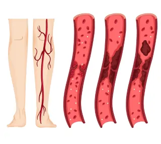
Deep vein thrombosis (DVT) is the formation of a blood clot in a ‘deep’ vein, commonly located in the legs or pelvis. DVT can cause serious complications, such as pulmonary embolism, if a blood clot (thrombus) breaks off and travels through the blood stream to lodge itself in the arteries of the lungs.
These blood clots are commonly found in the leg (calf), thigh (femoral), or pelvis (iliac). DVT is a type of venous thromboembolism, which is the third leading cardiovascular cause of death after heart attack and stroke. In India, there are approximately 2 instances of DVT, for every 1000 people. While this figure is lower than instances in Western countries, it is important to note that rural data is limited. (More detailed statistics further in the article).
The goal of treatment is to prevent the progression of these blood clots, the breaking loose, and development of complications such as post-thrombotic syndrome (PTS). Standard care begins with anticoagulants, with advanced options as the case profile intensifies.
Mechanism: How Does DVT Occur?
Think of DVT like a traffic jam in your bloodstream. Normally blood flows back to the heart seamlessly, but certain triggers make it pool, thicken and clot. This tends to happen in veins that are located deep inside the leg — typically the calf or thigh — hence the term Deep Vein Thrombosis.
DVT revolves around three basic things that occur simultaneously, known as Virchow’s Triad. These are stagnant blood flow, damaged veins, and sticky blood. For instance, if you sit in an airplane for hours, your blood will not flow as well, causing it to pool in your legs. Surgery or injury can also damage veins. If someone is pregnant, overweight, has cancer or takes birth control pills, their blood might be more ‘sticky’. Medically, ‘sticky blood’ is a hypercoagulative state or thrombophilia — the clotting system is overactive; platelets and proteins clump up too readily instead of flowing smoothly. If someone is a smoker, over 60 or has a family history of clotting (Factor V Leiden), that increases clotting risk too.
In the DVT context, this type of sticky blood pairs with slow flow or vein injury (Virchow’s Triad), to form deep vein clots faster. If a patient is recovering from knee surgery, bedridden or travelling for long (without moving around), the clot risks breaking off and travelling through the blood stream and lodging itself elsewhere. The most common and important of these risks is pulmonary embolism (PE), where the clot lodges itself in the lungs and blocks a pulmonary artery causing breathlessness, chest pain and in extreme cases, death. This occurs in 30-50% of untreated proximal DVT cases.
Conditions Treated
Obviously, this article is focused on the treatment methods and approaches for DVT. Within that condition, there are variations. Largely, the condition is based on the formation of clots in deeply located veins of the body that carry blood towards the heart. However, there are variations such as proximal DVT — usually in the upper thigh, such as the femoral or iliac veins — and distal DVT — in the lower calf. Sometimes, clots also form in the pelvic area, are and even the large inferior vena cava in the abdomen. All these fall under the larger umbrella of DVT.
Treatment largely revolves around two aspects — ensuring the clot does not become any larger and break loose. Without treatment, up to 50% of DVT occurring in the legs can lead to post-thrombotic syndrome, which is a long-term condition characterized by chronic pain, swelling, skin changes and sometimes, the formation of ulcers that do not heal easily. At a surface level, these symptoms mimic varicose veins, except DVT is not ‘visible’ like varicosities are.
Treatment also prevents the clot from recurring and preserves vein health, contributing to easier mobility. Wherever the clot has been diagnosed using an ultrasound scan — in the leg, arm, or elsewhere in the body — the early treatment with blood thinners, compression and other measures reduce greater risks.
Indications for DVT treatment:
The first thing to look for is severe pain in the leg; this could be accompanied by significant swelling and even discolouration of the skin. All these are signs that the leg needs to be addressed.
An ultrasound scan is the gold standard of diagnosis for acute DVT. Treatment is immediate, especially if it is proximal DVT, where the clots are located in the veins of the upper leg, such as the femoral (thigh) or iliac (groin) veins. When these veins are involved it’s also referred to as iliofemoral DVT.
In most cases, the first line of intervention is anticoagulant therapy, such as blood thinners given through an injection or taken orally. The more advanced treatments are used for severe cases of phlegmasia cerula dolens — where the leg becomes greatly swollen, turns blue and shows signs of tissue death from blockage of blood flow.
If the initial blood thinners have not worked within 24-48 hours, or if the patient is under 65 with iliofemoral DVT and low bleeding risk (no recent ulcers or major bleeds), it’s a reason for escalation. Catheter-directed thrombolytic therapy can also dissolve the clots quickly. These decisions are taken based on age, symptoms and comorbidities.
Step-by-step Guide to DVT Treatment
First, doctors confirm DVT with a duplex ultrasound — a painless scan showing blood flow and clots — and start blood thinners right away. The patient gets an IV heparin drip in hospital or a low-molecular weight heparin shot like enoxaparin under the skin for quick action.
Then, move to convenient daily pills: DOACs (Direct Oral Anticoagulants) are favoured over warfarin which requires regular blood tests (INR). These are planned for 3 months typically, but longer if risks persist.
The third phase is about helping the healing process by keeping the leg raised above heart level while resting (foot elevation pillows work well for this, as they do for pitting edema), wearing compression stockings (20 to 30mmHg) to reduce swelling, and walking small distances depending on the level of pain. Ideally, movement is medicine! This means bed rest, no matter how tempting, should be avoided. If swelling lingers after 48 hours, more advanced measures like clot-busting may be considered.
Finally, the patient must return for an ultrasound at 3-6 months to monitor the veins; therapy extends (6-12 months or lifelong) based on the cause of clotting, like cancer or genetics, to prevent repeats.
Advances in DVT Treatment
If standard blood thinners aren’t effective enough for acute iliofemoral DVT (clot extending from the groin to the thigh), doctors may recommend catheter-directed thrombolysis (CDT). A thin tube (catheter) is inserted through a tiny cut in a vein, and using real-time X-ray and/or ultrasound, it’s guided through the vein and placed directly into the clot, where it releases small amounts of clot-busting medicine, such as rt-PA, directly into the clot, dissolving it faster than pills alone. Often special devices such as AngioJet are used, which sucks out fragments while spraying the drug, which helps dissolve the clot faster — usually within 1-2 days.
Sometimes, doctors may include procedures like angioplasty (ballooning to open narrow veins) or stenting to improve long-term flow. CDT relieves severe pain and swelling sooner, and may relieve symptoms of post-thrombotic syndrome (PTS), such as chronic leg heaviness, although it does not prevent it entirely.
For those unable to be on blood thinners (for example, bleeding risks), an IVC filter, which is a small umbrella-like device, is placed in the vena cava, catching blood clots that are on their way to the lungs. These are minimally invasive, help you move sooner, and have few side effects such as bleeding and movement of the filter. The specifics are determined by doctors, based on patient profile.
DVT & VTE Treatment in India: Key Statistics
DVT treatment in India shows high success rates, with 80-95% clot resolutions in urban centers, however, rural data is sparse.
Incidences in India:
Among the general population, there are approximately 2 instances for every 1000 people. VTE in hospitals is at 17-22 per 10,000 admissions.
Post-orthopedic surgeries, DVT is a typical risk. 58% of DVT cases come from TKR (Total Knee Replacement), 26% of cases come from THR (Total Hip Replacement), with 6% of cases being proximal DVT. Hip fracture is responsible for 42% of cases (7% of these are proximal DVT).
Surgical patients at risk: 50-60%, medical patients are at 40-45%. Outcomes: 1 month VTE/mortality post-surgery is at 1.5%. Cancer patients have 2-20% VTE incidence. Recurrence is at 20-25% in 5 years, PE (Pulmonary Embolism) presents in 13-30% of VTE cases. Around 80% of PE cases can be fatal.
Outcomes:
1 month VTE/mortality post-surgery is at 1.5%. Cancer patients have 2-20% VTE incidence. Recurrence is at 20-25% in 5 years, PE (Pulmonary Embolism) presents in 13-30% of VTE cases. Around 80% of PE cases can be fatal.
| Metric | India | Western Countries (e.g., US/Europe) |
|---|---|---|
| General population (per 1,000) | 1.79 | 1 - 2 per 1,000 annually |
| Hospital VTE (per 10,000 admissions) | 7.46 - 17.4 | 20 - 25 |
Benefits of DVT Treatment
In short, it could be life-saving. A clot that embolizes in the pulmonary artery can cause pulmonary embolism, which could be fatal if untreated.
Treatment also reduces recurrent venous thromboembolism (VTE) by over 90% with blood thinners, and curbs post-thrombotic syndrome (PTS) — advanced CDT can reduce PTS by as much as 15% in clinical trials (ATTRACT trials), reducing leg pain and swelling long-term.
Up to 50% of untreated proximal DVT cases develop PTS, but compression and clot removal drop severe PTS by 25-30%. Advanced CDT can halve leg pain/swelling scores long-term. The numbers overwhelmingly favour starting therapy quickly.
Once swelling reduces, the quality of life increases. Within weeks, regular work and life activities can be resumed. Veins stay more open, preventing chronic issues. DOACs like rivaroxaban simplify life: no frequent blood tests or dietary hassles like with warfarin — it’s reliable protection, with a worry-free recovery path.
Rehab and Recovery
Patients should expect to be on blood thinners for 3-6 months or longer, to prevent clot movement. As mentioned earlier, high-knee compression stockings with 20-30 mmHg pressure are to be worn daily; these squeeze the legs to prevent clotting, swelling and the risk of PTS. Also, elevation of legs above the heart level, several times a day, is recommended. Particularly when resting. A foot elevation pillow is recommended. This elevation drains fluid naturally.
The rehab process begins with simple measures such as ankle pumps, which involve pointing and flexing the toes 10 to 20 times each hour, and short walks to improve the flow of blood. Seated calf raises, and short walks are recommended to restart circulation without strain. Gradually, activities such as stationary bikes and swimming, which are very healthy for the joints, can be added to improve circulation and prevent PTS through the reduction of pain and ulcers.
Doctors/medical teams keep track of progress for two years, and as the swelling subsides, activities are returned to. Early rehab helps to significantly reduce hospital stays and improve productivity at work, while fast tracking returns to regular life. The key is listening to the body and being very consistent and intentional with the rehab process. Part of being intentional is to report any new sensation, especially pain.
Risks and Complications
Blood thinners have a high efficacy but also come with a small risk of bleeding, which is 1-3% for serious cases like GI bleed or nosebleeds. It’s important to be careful and watchful of easy bruising, blood in urine/stool, or unusually heavy menstrual bleeding. These should be reported immediately, as they can be corrected with dose adjustments or reversal agents.
Advanced catheter-directed thrombolysis (CDT) has an associated 1-5% major bleed risk, especially in patients over 65 years of age, although no brain bleeds have been reported in clinical trials. There have been very rare cases of vessel puncture bleeding or allergic reaction to dye used for visualization.
Other possible complications include infection at the injection sites (<1%), post-thrombotic syndrome (PTS) in 40-50% of patients despite treatment (causing chronic ache/swelling in the leg), and IVC filter problems such as clot formation on the filter (5-10%) or filter migration during removal. Doctors will screen for possible complications, monitor the patient, and weight benefits against the risks. Usually these are minimal, and patients come through with great outcomes with simple precautions like avoiding combat sports.
DVT Clot Recurrence Risk
Generally, there is a 20-25% chance within 5 years from the initial episode. This risk escalates to 40% if the condition was unprovoked or untreated.
Provoked DVT: There’s a 10-15% risk within 5 years, if DVT was caused by surgery.
Unprovoked/Idiopathic DVT: There is DVT that doesn’t occur post-procedure, or any outside interference with blood vessels. There’s a 30% risk of clot recurrence within 5 years; men have a 5-fold risk, compared to women.
Is there a lifetime risk? Not necessarily. Long-term anticoagulant therapy can prevent recurrence by more than 90%. If therapy is stopped, then the risk rises to 5-10% in the first year, and 1-3% annually thereafter. There are other factors too — obesity, immobility or thrombophilia — which compound this risk. Lifestyle changes can hugely reduce these risks, along with preventive checkups.
Doctors determine risk of recurrence through D-dimer or ultrasound tests to tailor therapy appropriately.
Specific to India, the recurrence is about 6% in surgical patients. In thrombophilia-positive patients, the recurrence is 17.5% in 2 years and 24.6% in 5 years.