

Arteriovenous Malformations
 Home
Home- Arteriovenous Malformations


Medically Reviewed By Dr. Antariksh Vijan Updated on August 18, 2026
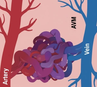
In the human body, arteries and veins function as independent networks of blood vessels with different functions. Typically, arteries carry oxygenated blood from the heart to the body, and veins carry de-oxygenated blood back to the heart. Hence, the ‘type’ of blood that flows in each is different. What happens if the two types of blood get mixed? This happens as the result of an arteriovenous malformation (AVM).
Normally, the two are connected through smaller, specific blood vessels called capillaries. In the case of AVMs, the arteries end up connecting directly to veins. Capillaries are the ‘buffer’ vessels responsible for delivering oxygen to the tissues. Only after this step can the deoxygenated blood enter the veins. With AVMs, this step is completely skipped, and the blood rushes almost too quickly from the arteries into the veins, resulting in a very low absorption of oxygen by the surrounding tissues. AVMs can develop anywhere in the body; however, there are certain regions where they are far more critical.
They’re found in the skin, lungs, spine or legs; however, brain AVMs are the most dangerous because the tangled vessels are weak and prone to bursting. If they do, it causes a hemorrhagic stroke — which can result in permanent brain damage, seizures or even death. Most people with AVMs are born with them, although the symptoms don’t show until adulthood. As such, there is no significant data on AVM occurrence in India, but global studies suggest an occurrence rate of 15–18 per 100,000 adults. However, the Cerebrovascular Society of India notes brain AVMs are rare.
Causes
Typically, AVMs form while developing as a fetus inside the mother’s womb. As the fetus grows, tiny chemical signals guide vascular formation — the mesh of arteries, capillaries, and veins forms as a result of these signals. Sometimes, the signals get disrupted, causing the arteries and veins to connect directly. This happens very randomly, like an electrical malfunction, and isn’t necessarily caused by anything the mother did during the pregnancy. That said, AVMs can sometimes occur after birth through injuries or radiation exposure. For instance, if someone is undergoing radiation therapy for cancer around the vicinity of their blood vessels, new abnormal connections may form in that region. Sometimes, trauma to tissues can also trigger AVM formations, but this is significantly less common than fetal AVMs.
Most AVMs are not inherited; however, certain genetic conditions can considerably increase the risks of developing malformations. Of these, the most important is hereditary hemorrhagic telangiectasia (HHT) or Osler-Weber-Rendu syndrome. People with HHT have a gene mutation that makes them more likely to develop AVMs in various organs. If one of the parents has HHT, then there’s a 50% chance a child will inherit the condition. However, the percentage of HHT cases that can reliably produce an AVM is still unclear. There are other rare genetic disorders that may also increase risk, but these are much less common. For most families, AVMs appear as a one-time event, very rarely showing up in future generations.
Risk Factors
As explained above, AVMs are fairly arbitrary. They appear without any clear or sustained risk patterns or family history. So, any categorization here is loosely based on collective studies and patterns found globally:
- Genetic standpoint: HHT is the biggest risk factor. If a parent has HHT, each child has a 50% chance of having it. In the very odd scenario that both parents have HHT, there is a near 100% chance of a child inheriting it. The field of genetic counselling has emerged in prominence to assess heritable factors and assist people with family planning, so the chances of transferring these conditions to future generations can be minimized.
- Gender: Males tend to have AVMs more often than females.
- Age: People born with AVMs do not experience any symptoms immediately. They tend to appear between the ages of 10 and 40 years. The ‘greatest’ risk period is between ages 30 and 50. Those older (> 60 years) have greater risks of bleeding if they have AVMs.
- Family history: This is a tangential risk. If many close relatives have HHT and/or multiple AVMs, then the risk is higher. That said, a majority of cases don’t involve any family history.
- Radiation treatment: Radiation treatment to areas near blood vessels can increase the risk of developing AVMs later. A previous AVM bleed also increases the risk of future bleeding.
Symptoms
AVMs often have no symptoms and are only detected if imaging tests are done for other medical reasons. There are many individuals who remain unaware of their AVM condition until bleeding or neurological complications arise. When symptoms do occur, they depend on the location. Since brain AVMs are the most critical, let’s examine the symptoms:
- Seizures (the most frequent symptom after bleeding)
- Intense headaches (occur suddenly, one-sided, or in the "worst headache of my life" category)
- Weakness, numbness or paralysis in one limb
- Loss of coordination and balance resulting in difficulty walking
- Vision disturbances (vision loss and eye movement disorders)
- Difficulty speaking or comprehending language
- Dizziness, nausea and vomiting
- Cognitive decline worsening over time, memory loss, confusion and dementia
- Hallucinations
- Back pain (this is more specific to Spinal AVMs)
In newborns (with a Vein of Galen malformation): an enlarged head, swollen scalp veins, seizures, failure to thrive, heart failure. Typically, seek care for a seizure, sudden disorientation or limb weakness, vision changes or a sudden severe headache — these are the most telling signs of an AVM that requires intervention.
Diagnosis
The first step is a physical exam where symptoms are reviewed closely. Doctors may first listen for a bruit — a ‘whooshing’ sound made by blood rushing through abnormal blood vessels. The next step is imaging:
- CT scan: This uses X-rays to detect the presence of bleeding and abnormal structures in the brain or spinal cord.
- MRI: An MRI shows more detailed tissue images, often catching smaller changes that other scans may miss.
- MRA (Magnetic Resonance Angiography): An MRI angiograph that focuses purely on blood vessels and blood flow patterns.
- Cerebral angiography (DSA): This is the gold standard. Here, a particular dye is injected in the artery and X-rays are taken in real time to study the blood vessels and reveal the AVM’s exact location and structure.
- Transcranial Doppler ultrasound: Uses sound waves to measure blood flow velocity through the vessels.
It’s worth noting that many AVMs are found incidentally during imaging for other concerns like headaches and head trauma injuries.
Treatment
Treatment methods for AVM are decided based on the size, location and bleeding that may have occurred.
- Conservative management involves observing the AVM without treatment. This is recommended in cases where there are small, unruptured AVMs in difficult-to-reach areas. Medications can help manage symptoms like headaches, back pains and seizures; however, they do not eliminate the AVM itself.
- Surgical resection (open surgery) is considered for small, accessible AVMs. This involves removing the entire AVM from the brain or spine with the aid of a microscope. The advantage is that the patient is considered fully cured post-removal. Of course, it’s a delicate surgery that does present its own risks.
- Endovascular embolization is less invasive. It involves the insertion of a catheter into blood vessels to inject glue or coils into the AVM to stop blood flow. This is also used as a pre-surgery procedure to improve surgical outcomes.
- Stereotactic radiosurgery (Gamma Knife) involves damaging the AVM vessels with radiation to make them close between 1 and 3 years. This approach is effective for small AVMs in deep and hard-to-reach areas.
- Multimodality treatment involves a combination of two methods — like embolization combined with surgery — to treat complex AVMs.
Benefits of Treatment
The primary objective and benefit of AVM treatment is the prevention of a hemorrhagic brain stroke. Once this is dealt with, the chances of further bleeding from that AVM are practically non-existent. Additionally, treatment frequently helps to reduce or stop seizures, which can be the most common and distressing symptom of brain AVMs. Treatment helps many patients with their symptoms of headache, weakness, numbness or other neurological patterns. There is also a huge psychological and guessing element to this.
The idea that at any arbitrary moment, a brain AVM can trigger a severe medical crisis is an extremely anxiety-inducing scenario to live with — for patients and their families. The worry of a sudden unexpected bleed escalating into a stroke or cognitive decline is very real. Successfully treated AVMs avoid this, and especially in children, they avoid developmental problems. In older adults (>60), an untreated AVM carries a high lifetime hemorrhage risk.
Recovery & Rehabilitation
The timelines for recovery and rehab vary according to the treatment. Some are simple outpatient procedures; others have a more specific layered road to full recovery.
Stage 1: Hospital recovery (Days 1–7)
- Embolization: This involves 1–3 days in the hospital. Same-day discharge is fairly common.
- Surgery: 4–6 days in total (one day in a neuro-ICU followed by a step-down unit).
- Radiosurgery: Same-day discharge; a patient can go home soon after the procedure.
Patients are advised to get out of bed within 24 hours, to avoid any complications.
Stage 2: Early home recovery (Weeks 1 to 6)
This stage involves a phased reintroduction to activities. Initially, heavy lifting or any form of strenuous exercise is prohibited. However, by week 4, a typical daily routine, including some form of physical training, can be resumed. For brain AVMs, driving is prohibited for 6 months due to the very delicate nature of the situation. Basic wound care protocol applies — keeping the wound dry and clean, and watching for signs of infection.
Stage 3: Complete recovery (Months 2 to 6)
Complete recovery takes anywhere between 2 to 6 months. In the case of radiosurgery, vessels close between 1 and 3 years. Most normal activities and exercises can be resumed around this time, including a timely reintroduction to resistance training. Rehabilitation:
Rehabilitation focuses on fine motor control, ambulation, balance and strength. Trunk stabilization is often a focal area. Occupational therapy may help with daily task planning and adaptive techniques. Speech therapy is introduced for those with language difficulties and swallowing problems. Evidence shows that children improve across all domains after inpatient rehab.
Long-term follow-ups involve MRIs after 5 years to confirm complete closure. Post-discharge, quarterly check-ups are the norm. Once the patient is considered stable, these are phased into bi-annual and annual check-ups.
Risks if Left Untreated
High-risk or symptomatic AVMs are not a casual ‘wait and watch’ affair. If left untreated, they pose very serious risks. These are weak blood vessels prone to bursting. In the brain, this could cause a hemorrhagic stroke which may cause permanent brain damage, paralysis, vision loss, speech problems or even death. The chances of rebleeding are very high with AVMs. In the first 20 years, the risk of a rebleed is 51%, compared to 33% among non-bleeding AVMs.
Untreated AVMs cause seizures in about 18% of patients during 20 years; this risk is much higher (44%) for younger patients aged 10–19. AVMs also deprive brain tissue of oxygen, since blood bypasses the capillaries. This leads to gradual weakness, cognitive decline and dementia over time. Diagnosis can be challenging because these gradual changes mimic other conditions. Older adults (>60 years) face an 89% hemorrhage risk over 9 years. In very severe cases, bleeding can cause brain death; this accounts for about two-thirds of all fatalities related to untreated AVM patients.

