

EVAR Procedure
 Home
Home- EVAR Procedure


Medically Reviewed By Dr. Karan Anandpara Updated on August 10, 2026
Endovascular Aortic Repair, commonly referred to as EVAR, is a minimally invasive procedure used to treat abdominal and some thoracic aneurysms. An aneurysm is a weak, stretched out blood vessel that has ballooned outwards like a bulge. This is due to this portion of the wall becoming thin. It has lost the healthy springiness of a regular vessel. However, there are no symptoms indicating its existence, which makes the situation more precarious. An aneurysm can tear or burst, leading to sudden internal bleeding and life-threatening emergencies.
EVAR is a stent-graft procedure increasingly adopted for complex and ruptured aneurysms. Generally speaking, it is favoured for high-risk patients who are not good candidates for invasive surgery. They may be older or have pre-existing heart and lung issues. As a result, they may not be able to tolerate major surgery.
As a very broad rule, doctors tend to favour surgical resolution of a problem as it provides full access to the site. However, this is only a textbook ideal. Actual patients come with a variety of comorbidities that make surgical resolution and treatment protocols entirely dependent on the individual risks. This nuance drives a lot of medical research.
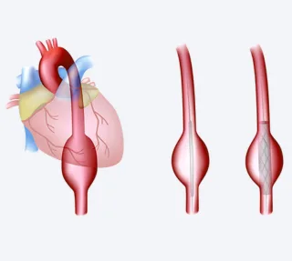
EVAR tends to be considered for patients who are older or those with pre-existing heart and lung issues. They may not be able to tolerate major surgery, as a result, a minimally invasive procedure (though more complex, in some senses) is considered. Doctors guide a small tube like stent-graft through the groin, into the aorta. This is a sort of bypass — it creates a newer channel for the blood to flow, taking pressure off the weakened bulge and lowering the risk of a life-threatening rupture.
In India, EVAR is mostly concentrated in super speciality, high-volume cardiac care centers with hybrid ORs and advanced cardiovascular care. As a result, comprehensive national data via a registry is hard to come by. That said, center-wise reports are incredibly encouraging, and match global success rates of 95-98% with reintervention rates of 10-2% over 5-10 years. Aneurysm related mortality itself remains low.
Conditions Treated with EVAR Procedure
The primary condition addressed by EVAR is abdominal aortic aneurysm (AAA). It is also used to treat aneurysms in the chest portion of the aorta; in those cases the procedure is referred to as TEVAR.
The objective of an EVAR is to exclude the diseased portion of a vessel, and create an inner tube/newer channel, so blood circulation to limbs and vital organs is maintained.
While evaluating a patient for EVAR, doctors consider the rate size of the bulge and the rate at which it is expanding. The more it expands, the closer it gets to a rupture/tear. For an infrarenal abdominal aortic aneurysm anything greater than 5.0-5.5 cm or rapidly enlarging at the rate of 0.5 cm/6 months can warrant an EVAR.
Aside from these primary conditions, the procedure is also used to address penetrating aortic ulcers, pseudoaneurysms, traumatic aortic injury, and some chronic post-dissection aneurysmal dilatations, depending on anatomy.
Indications and Contraindications for EVAR Procedure
In simple terms, EVAR is considered when the risk of an aneurysm rupturing is greater than the risk of the procedure itself. Essentially, it’s a question of risk assessment. The second factor is whether the anatomy itself makes the procedure feasible.
Symptomatic AAA is a consideration, even if the size of the aneurysm is slightly below the standard level (>5.5cm in men, >5.0cm in women), when new symptoms start appearing — back, abdominal or flank pain.
Asymptomatic AAA is an indication, if the aneurysms are above standard levels, and at risk of rupture. However, detection is the issue. And they often come to light by ‘accident’, during standard preventive health checkups — which further underlines the need for these. A CT, MRI or ultrasound done for another reason can incidentally detect these. In older men, especially smokers or people with a family history (again, unpredictable), a screening ultrasound can pick details.
Sometimes, a doctor can feel a ‘pulsating’ mass in the abdomen, although this manner of physical examination and follow-up detection is a bit unreliable. Most AAAs are discovered through targeted abdominal ultrasounds, and the detection often happens as a result of another issue — back pain, bowel problems, kidney issues or something unrelated.
A ruptured AAA obviously merits an EVAR. It can be life-saving and is preferred to open surgery, with lower mortality rates. However, this depends on the center having rapid endovascular access. In patients with lots of comorbidities — coronary artery disease, diabetes, chronic obstructive pulmonary disease (COPD), renal issues — open surgeries automatically come with prohibitive risks. Here, EVAR is a preferred approach.
Contraindications for EVAR include blood vessels in the neck that are extremely tortuous (curved or unusually twisted), heavy calcification or damage at the landing zone (the part of the artery where the stent graft is meant to grip and seal) or the inability to maintain lifelong imaging.
If the landing zone is too short, bent, highly calcified (hard), or fully clotted, then the graft won’t seal well, leading to leakage issues. EVAR works best with regular post-procedural followups, because the repairs change over time. A graft can shift, leak or the aneurysm sac can start filling again. Imaging may not be possible for various reasons, including access, costs and sometimes the feeling of ‘doing okay’ after the procedure.
Reference: European Journal of Vascular and Endovascular Surgery – ESVS 2024 Clinical Practice Guidelines on the Management of Abdominal Aorto-Iliac Artery Aneurysms. EjvesEndovascular Today
Step-by-step Guide to EVAR Procedure
The first pre-procedural step is a detailed CT angiography of the aorta from the chest to the femoral muscles. This is absolutely crucial in presenting a more detailed picture of the anatomy including vessel size, aneurysm size and location, neck length, and vessel health/calibre.
After this, comes device planning. Vascular & interventional radiologist have to select the stent-graft according to their findings. Diameter oversizing is an aspect of this — doctors usually choose a device that’s 10-20% above the native aortic diameter. Other aspects include length to cover aneurysm, and modular components for iliac limbs.
Then, comes risk stratification and optimization for comorbidities. This involves selecting and detailing doses of medications to ensure compromised organ systems aren’t adversely affected — cardiac evaluation, renal protection strategy (intra and post-op), antiplatelet and anticoagulant strategy, and anesthetic assessment.
The actual EVAR procedure itself can be broken into three stages:
Access to groin arteries — This involves puncturing the groin arteries under the regional or general anesthesia (type of anesthesia varies by patient profile). These ‘punctures’ are actually small and precise incisions in the groin arteries or sometimes, percutaneous punctures. The percutaneous puncture is less invasive than the groin incisions, but is very dependent on vessel anatomy and can fail in some patients, in which case, incisions are a plan B.
After this, guidewires and sheaths are advanced towards the target area using a live x-ray (fluoroscopy) for guidance.
Then, a large delivery catheter containing the compressed stent graft is advanced through the iliac arteries into the aorta, across the aneurysm. The main body is positioned just below the renal arteries. Once this is done, the stent-graft is released and expanded to seal against the healthy aortic wall. Iliac limb components are then deployed to connect the main body to each iliac artery, which successfully diverts blood away from the aneurysm sac. This is the objective of the procedure. Balloon molding may be performed to improve the seal and fixate the graft.
Once finished, a completion angiography checks for leaks (residual blood flow into the aneurysm sac), correct positioning and patency of branch vessels. The sheaths are then removed, access sites sealed (using sutures or closure devices). Skin incisions may be covered with dressings without any large stitches.
Reference: Journal of Vascular Surgery – The Society for Vascular Surgery Practice Guidelines on the Care of Patients with an Abdominal Aortic Aneurysm (2018).
Advanced Techniques in EVAR Procedure
These techniques are used when the aneurysm is located in a very difficult place or the artery shape is not ideal for standard repair.
- Fenestrated EVAR or FEVAR is a technique that has small openings to keep blood flowing to the kidneys and other vital branches. The grafts themselves are made with small holes to facilitate this.
- Branched EVAR or BEVAR involves stent grafts with side branches for visceral arteries. This is used to treat extensive and highly complex aneurysms in the thoraco-abdominal space. It’s a very technically demanding procedure, often limited to super speciality/high-volume centers.
- There is also PEVAR or percutaneous EVAR where, as explained in the previous section, the device is placed using tiny skin punctures instead of a groin cut. Preclose techniques help seal the artery afterwards.
- There are also intra-operative advances like chimney or snorkel techniques that use parallel covered stents placed into visceral arteries, alongside a standard aortic graft. This is resorted to when FEVAR is not possible or in urgent settings.
- Another area of advancement is the management of endoleaks — residual leakage into the aneurysm sac, even after the procedure. Coils, plugs, glues or even additional cuffs/limbs are used to treat endoleaks. Sac embolization is also used for persistent leakage.
Recovery, Follow up and Rehabilitation
The most important thing about the recovery process is lifelong surveillance. EVAR recovery itself is not very tedious, but lifelong followups are essential to know that the graft is working. It’s a dynamic situation that can change, and should it, doctors take a call on how to fix leaks or other problems.
Immediate recovery begins in the hospital. There’s early mobilization work, once the hemodynamics are stable. Hospital stay is around 2-3 days. Once the anesthesia wears off, an oral diet can be resumed.
Most patients return to light activities within 2-4 weeks, and resume all their activities within 4-6 weeks. In the first month, heavy lifting (>10kgs) and high-impact exercises are discouraged. Light aerobic work – walking and cycling — is recommended.
At 1 month, a CT Angiogram or contrast-enhanced ultrasound is highly recommended. This is then repeated at 6-12 months, and thereafter on an annual basis. These follow ups check for endoleaks, graft mitigation, sac size changes and limb patency.
Despite a successful procedure, late complications can occur (e.g., type I endoleak, graft mitigation, limb occlusion). If these aren’t dealt with, the procedure itself becomes futile.
Structured rehab largely models itself on cardiac rehab programs with some changes. The usual rules always apply — controlling diabetes, stopping smoking, and managing blood pressure to avoid further vascular events.
Benefits of EVAR Procedure
Simply put, EVAR can save lives. Aneurysms can burst at any point, and there’s still no way of successfully predicting when. This makes the matter all the more dynamic, complex and scary.
EVAR is also minimally invasive, so recovery times are significantly less compared to open surgery. It is often the most feasible alternative for high-risk patients (older age, fragile, cardiopulmonary comorbidities).
However, it’s not a ‘one and done’ type solution. EVARs require lifelong monitoring and constant follow-up imaging. Surprisingly, this becomes difficult for a lot of patients who don’t live close to super-speciality centres. It also has a higher rate of secondary interventions, compared to open surgeries.
As with any procedure, there is rarely a clear winner. Doctors make decisions based on the best relative outcome. For many, EVAR is a far more gentle and high quality alternative to surgery.
Reference: The Lancet – Endovascular versus Open Repair of Abdominal Aortic Aneurysm in 15 Years' Follow-up of the UK EVAR Trial 1 (2016).
Risks and Complications of EVAR Procedure
Considering the delicate and unpredictable nature of aneurysms, the complications of EVAR are very carefully explained to patients and their families/caregivers. It’s crucial to understand these, before having the procedure as EVAR involves a lifelong commitment to follow ups.
Access site injuries — hematoma, pseudoaneurysms, femoral artery injury or dissection, especially in calcified and small vessels.
Thromboembolic events — limb ischemia (lack of blood flow), distal embolization or even stroke (more relevant in thoracic procedures).
Endoleaks — The main complication, which necessitates lifelong followups. Think of EVAR like a plumbing issue; fixing it once doesn’t guarantee that it remains okay for life. Pipes may clog, taps may leak; the plumbing will need follow up maintenance. Persistent blood leaks into the aneurysm sac can occur. These are classified based on their extent: type 1 (inadequate seal), type 2 (retrograde flow), type 3 (graft component failure), type 4 (porosity) and type 5 (endotension). An endoleak must be addressed to avoid future complications.
Graft-related — These are device-related. Migration, kinking, limb occlusion, device fracture or even infection (rare, but serious).
Systemic — These are the systemic risks. DVT (Deep Vein Thrombosis), kidney injury (from contrast dye), allergic reactions, myocardial infarction and very rarely paraplegia (limb paralysis) if thoraco-abdominal coverage compromises spinal perfusion.
The red-flags symptoms that patients and caregivers must look out for are: sudden back pain, sudden abdominal pain, syncope, leg pain or ‘coldness’, fever, or groin redness. Any of these merit urgent evaluation.

