

Left Ventricular Assist Device (LVAD)
 Home
Home- Left Ventricular Assist Device (LVAD)

Medically Reviewed By Dr. Vishal Pingle, Cardiothoracic & Heart Transplant Surgeon. Updated on May 12, 2026
LVAD treat end-stage heart failure by mechanically supporting the left ventricle's pumping function when medications and other therapies fail. If the heart is severely weakened due to end-stage heart failure, there are feelings of breathlessness, swelling, and exhaustion despite maximum medications. Then, an Left Ventricular Assist Device comes into play as an auxiliary heart pump. It performs most of the functions of the left ventricle, which helps pump blood to other parts of the body. Doctors use it in three main ways: As a "bridge to transplant" while a patient wait for a donor heart (common in India due to donor shortages); "bridge to recovery" if the heart might heal after rest; or "destination therapy", as a permanent fix if transplants aren't an option, like for older patients or those with other health issues.
Top centers in India report one-year survival rates of 80-90%. These outcomes match global standards. Many patients can even resume light activities, travel and begin rehab, thereby enjoying a better quality of life for years.
Conditions Treated by Left Ventricular Assist Device (LVAD)
Left Ventricular Assist Device's are mostly useful for patients suffering from advanced-stage heart failure (Stage D). This stage usually involves either ischemic heart disease, cardiomyopathy, or post heart-attack damage. The LVAD can be used to treat patients with predominant LV dysfunction, and at risk of irreversible organ damage.
Before going into the causes, here’s what the symptoms of Stage D heart failure look like: Swelling worsens in legs, ankles, abdomen, or lungs, causing rapid weight gain from fluid buildup and a bloated feeling. The patient may cough up frothy pink mucus, wake gasping at night (paroxysmal nocturnal dyspnea), or struggle to lie flat without propping up. Other signs include dizziness/fainting from low blood flow, irregular heartbeats, poor appetite, nausea, confusion, and kidney/liver strain leading to less urine or jaundice. These are all signs of slowly failing organs, due to LV dysfunction.
Common underlying causes:
- Dilated cardiomyopathy: Heart muscle stretches and weakens (often from past infections/viruses).
- Ischemic heart disease: Damage from blocked arteries or prior heart attacks.
- Valvular issues: Leaky or narrow valves exacerbating pump failure.
- Post-heart attack scarring: Left ventricle can't contract properly, leading to lowered cardiac output.
In India, these causes affect millions due to rising diabetes (which can mask symptoms), hypertension, and delayed care; LVADs step in when options like CRT (pacemakers) fail. Think of the interventions in stages:
- Level 1 (Early): Use of medications/devices to manage mild failure.
- Level 2 (Intermediate): Inotropes via IV for a temporary bridge.
- Level 3 (Late): LVAD for patients requiring continuous assistance.
Indications for Left Ventricular Assist Device (LVAD) Implantation
LVADs come into the conversation when the heart’s ejection fraction goes below 25%. At this stage, the heart is too weak to pump the required amount of blood to keep other organs functioning in a healthy manner. The risk of permanent organ damage and frequent hospitalizations looms large. This is Stage D (late stage) heart failure.
Key indications in patient tiers:
- Tier 1: Bridge-to-Transplant — A patient is on the donor waitlist but crashing fast; LVAD is a temporary solution, used commonly in India, due to longer waiting periods.
- Tier 2: Bridge-to-Recovery — Temporary support after a heart attack or viral infection to give the heart a better chance to heal.
- Tier 3: Destination Therapy — Permanent choice if transplantation is not possible due to age (over 65 years old), diabetes, kidney disease; NYHA (New York Heart Association) Class IIIb-IV symptoms — symptoms like breathlessness hit with minimal effort (IIIB) to appearing even at rest (IV) — for 45+ days despite optimal care.
- Tier 4: Bridge-to-Decision — For critically ill patients needing time to assess eligibility.
Types of Left Ventricular Assist Device (LVADs)
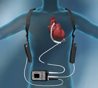
There are various types of left ventricular assist device based on the requirements of the patient — how long they may need support and the specific condition of the heart. The pump is located inside the chest and is attached to an external battery that can be carried in a pouch or backpack.
- Continuous flow LVAD (most common type today): Uses impellers like a fan to maintain continuous flow of blood and is relatively quiet and smaller compared to previous LVAD devices.
- HeartMate 3: Latest model with magnetic levitation to cut clots; great for long-term use (bridge or permanent). Over 40,000 implanted worldwide.
- HeartMate II: Axial flow, reliable for bridge-to-transplant.
- HVAD (less common now): Centrifugal flow and compact in size.
- Pulsatile flow pumps (first-generation, infrequently used): Replicate your body’s inherent “thump” using filling/squeezing chambers; larger, greater chance of infection.
Specialized types:
- RVAD/BiVAD: Right or both ventricles if needed.
- Pediatric: Smaller for kids.
In India, HeartMate 3 dominates, due to durability (80-90% 1 year survival outcomes) and a significantly lower risk of strokes, bleeding and other types of events. There are Indian projects like the Hridayantra which focus on cost-effective indigenous LVADs. However, these are still in the R&D stage, and yet to reflect in any clinical trials.
Step-by-Step Guide to Left Ventricular Assist Device (LVAD) Implantation
Implantation of Left Ventricular Assist Device is a complex open-heart or minimally invasive surgery procedure that takes 4-6 hours, where surgeons insert a mechanical pump inside the chest to support a malfunctioning left ventricle to circulate blood. The procedure is performed under general anaesthesia. Here’s a stage-wise breakdown of the process:
Step 1: Preparation (1-2 hours)
Complete medical assessments, establish IV lines, antibiotic infusion, and assembly and sterilization of the LVAD pump. The patient is hooked up to a heart lung machine that takes over circulation.
Step 2: Access and Core (1 hour)
Surgeons make an incision across your breastbone (sternotomy) or mini-incisions (thoracotomies, in minimally invasive procedures), access the heart, stitch a ring (cuff) at the tip of the left ventricle, and harvest a piece of heart muscle using an instrument. Echo guides precise placement.
Stage 3: Connect & Test (1-2 hours)
Insert inflow tube into ventricle, attach outflow graft to aorta, tunnel the power cable (driveline) through abdominal skin, connect controller/batteries, and test pump flow before closing chest.
Stage 4: Close & ICU
Stitch up, move to ICU for 3-7 days of monitoring as the pump ramps up.
While traditional open-heart pump insertion provides for greater visibility, minimally invasive approaches are catching up — matching success rates with 50% less blood loss, fewer days in the ICU, reduced infection risk and lesser strain on the right side of the heart. However, eligibility for a minimally invasive implantation is dependent on coronary anatomy, prior surgical history and a variety of other factors, which doctors first assess before deciding.
Benefits of LVAD Implantation
LVADs offer a transformational experience for end-stage heart failure patients, helping them regain some control, when the heart can no longer cope. Here is a phased perspective on what lies ahead:
Short-term (first weeks): Immediate relief from breathlessness and fatigue as the pump takes over pumping — the kidneys/liver recover, swelling drops, and constant hospital stays are avoided. Many feel energy return within days, starting light rehab in ICU.
Medium-term (1-6 months): Participation in everyday activities such as walking, cooking and driving becomes possible. Quality of life significantly improves—with 80% reporting fewer symptoms compared to taking medications alone.
Long-Term (1+ Years): Up to 5-7 Years Survival (58% with HeartMate 3). Some patients wean themselves if their heart function improves. Travel, work, and family time become possible with portable batteries; many resume their hobbies. Destination therapy is ideal for non-transplant recipients.
Overall, LVADs cut mortality risk by 50% vs. drugs, boost exercise capacity, and improve mood.
Risks of Left Ventricular Assist Device (LVAD)
LVAD surgery and their lifelong use entail certain dangers; however, with contemporary LVADs such as the HeartMate 3 and proper medical care from hospitals, most of the dangers have been substantially reduced to 20-10%. Here are the risks categorized:
Early Postoperative Complications (within 30 days, 10-20% likelihood):
- Bleeding due to anticoagulants or use of cardiopulmonary bypass (the most frequent complication in early stages).
- Right heart failure (15-25%), requiring temporary medication or management.
- Infection around the surgical incision or driveline site; strokes caused by clots or air bubbles (7-11%).
Complications Related to Device or Pump Function:
- Thrombus formation within the pump (less than 2% with HeartMate 3 compared to earlier devices).
- Malfunctioning (very rare, 1-2%/year); leaking aortic valve due to continuous flow.
Long-term Risks (within 30 to 50%, over several years):
- Infection (driveline, 60% within 90 days) that is treatable with antibiotics but dangerous.
- Bleeding (GI/nose area, 20 to 30%) because of thinners plus pump effect.
- Strokes (ischemic/hemorrhagic, 10-15%); kidney/liver strain.
A huge part of risk management is thorough risk profiling, prior to the surgery. For this, the heart team takes stock of various comorbidities — age, diabetes, and more. Many of these are manageable with monitoring, hygiene and meds.
Recovery and Rehab
The LVAD patient recovery process is divided into 3 stages and enables the patient to gain strength in a safe manner while also learning how to handle the device.
Stage 1: In-patient period (days 1-21)
ICU admission (3-7 days): Weaning off the breathing tube shortly and beginning with some light walking and sitting under PT supervision (to prevent clotting). Learning alarms, battery changes, driveline maintenance. Transitioning to step down unit.
Stage 2: Early post-op outpatient (weeks 4-12)
Discharge with outpatient follow-up care: Cardiac rehabilitation 3-4 days per week including exercise (treadmill/bike at low to moderate intensity 20-40 minutes), emphasizing stair climbing and dressing. Most patients experience “normal energy” by 3 months. No driving, water submergence as yet.
Stage 3: Maintenance period (>3 months)
Full activities can be resumed after discharge such as work and other recreational activities (for 80-90%). Continuing with physical therapy and weight training along with flow monitoring (4-6 liters per minute). 80-90% of Indian patients hit milestones matching global data.