

T-TEER Procedure for Tricuspid Valve
 Home
Home- T-TEER Procedure for Tricuspid Valve


Medically Reviewed By Dr. Meghav Shah Updated on August 18, 2026
Tricuspid valve repair addresses regurgitation of the tricuspid valve, which regulates the flow of oxygen-poor blood to the lungs. The tricuspid valve is located between the right atrium and the right ventricle. Blood that passes through it goes from the right atrium to the right ventricle, and then, the right ventricle pumps it into the lungs (via the pulmonary artery) for oxygenation. In the broader scheme of things, tricuspid valve repair addresses valve regurgitation, tricuspid stenosis, congenital (by birth) lesions, RV dysfunction, symptomatic right-sided heart failure and a few other conditions.
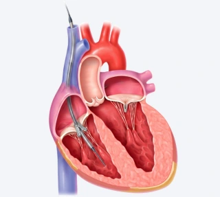
For the purposes of this article, we will look at the application of TEER (Transcatheter Edge-to-Edge Repair) in tricuspid valve repair. It is a minimally invasive way of treating symptomatic regurgitation in the tricuspid valve. The principle of TEER is to clip leaflets of valves to prevent regurgitation. Since it is done without opening the chest surgically, it is usually recommended for frail, high-risk, older patients. While TEER as a methodology has been more extensively adopted for mitral valve regurgitation, it is increasingly being done for tricuspid valve regurgitation too. TEER for tricuspid regurgitation is commonly referred to as T-TEER.
In India, extensive data about annual caseloads are not yet reliably published. However, single-center studies show success outcomes matching global rates. Global studies show a reduction in regurgitation to 'moderate' or 'less' within 30 days and only a 3% rate of major adverse events. Multiple trials - CLASP TR (which monitored outcomes of the PASCAL system), TriClip and EVOQUE — all showed very meaningful outcomes in terms of reduction and improved quality of life.
Reference: Donal E, Dreyfus J, Leurent G, et al. Transcatheter Edge-to-Edge Repair for Severe Isolated Tricuspid Regurgitation: The Tri.Fr Randomized Clinical Trial. JAMA. 2025;333(2):124–132. doi:10.1001/jama.2024.21189.
Conditions Treated
T-TEER is primarily used to address severe symptomatic tricuspid regurgitation (TR). This could be moderate to severe TR, especially functional TR caused by atrial fibrillation, right-side heart enlargement, pulmonary hypertension, or other valve-specific diseases. TR is considered severe when the valve is leaking enough to cause breathlessness, fatigue, leg swelling, abdominal bloating or fluid build up in the lungs. Usually, this is caused by damage to the valve itself.
On the other hand, functional TR means there is no damage to the valve itself, but similar effects — this can be due to an enlargement of the right atrium or right ventricle, which pulls the valve ring apart, and prevents the leaflets from connecting properly. Hence, ‘functionally’ the same as a regular TR.
The first line of intervention is almost always medical therapy. T-TEER is considered when symptoms persist despite that. Especially in people who are not good candidates for open-heartmitral valve surgery — due to age, frailty, or a high surgical risk profile due to comorbidities.
Before recommending T-TEER, doctors look at the heart’s pumping strength. People with a relatively decent RV function and without severe lung pressure problems are likely to benefit. RV function is measured via an echo and a preserved right-ventricular function is a TAPSE around 19mm and RVFAC above 46% before the procedure. An RVFAC > 35% is also considered workable. Anything below 30% is treated as RV dysfunction — in this case, the risks of a T-TEER may outweigh the benefits.
Step-by-step Guide to T-TEER Procedure
The first step is a diagnostic evaluation. An echocardiography confirms that the regurgitation is significant enough to warrant intervention. Next comes a transesophageal echo which gives doctors a better understanding of valve anatomy — leaflet shape, size of the leak and coaptation gap.
A heart team reviews the profile to decide between TEER or other interventions, based on the anatomy and overall risk profile of the patient. A specific assessment of the right-heart shows if the right ventricular function is still good enough to benefit from the procedure. Not all cases are a yes or no. Many sit in between, and the discretion is left to the medical teams upon consideration of many other factors.
Very large leaflet gaps, very distorted valves or extremely low right-ventricular function can make TEER less feasible. A global study showed that of patients considered for T-TEER, about 20% were clearly favourable, 48% were feasible, and about 32% were unsuitable because of anatomy.
Once the procedure has been decided upon, there are a few clear stages:
- Anesthesia is given and an ultrasound/echo helps map the valve and plan the best angle for clipping.
- Then, a small tube (catheter) is inserted through a groin vein and guided towards the right side of the heart.
- Next comes the tricky part — positioning the clip device across the tricuspid valve while using real-time imaging.
- As explained earlier, the objective is to bring the leaflets together to reduce regurgitation.
- Once the position looks right, the leaflets are grasped and gently pulled together.
- The clip is closed to test how much the leakage has been reduced.
- If the reduction is not satisfactory, another clip may be added and testing is done again.
- Once the result is confirmed, the clipping device is ‘released’, the catheter is withdrawn and the puncture site is closed.
- For a short time, patients are monitored with follow up echo scans to check the valve and recovery. In uncomplicated cases, a discharge may be possible on the next day. However, these protocols vary slightly from team to team.
Advanced Techniques in T-TEER Procedure
Multiple clipping is one of the most crucial advances. Two or more clips may be placed at a combination of angles to close the leak sufficiently. In some patients, something called the ‘clover technique’ is employed. This means creating more than one smaller opening instead of attempting to close the entire leak with a single clip.
This strategy is employed when the valve gap is so wide that conventional T-TEER won’t work. The presence of a pacemaker is a crucial consideration here. If one is present, the doctor must weave around the pacemaker wire very carefully to avoid trapping or disturbing it during the procedure.
Advanced imaging is a huge part of the story.
3D echocardiography can offer a three dimensional view of the valve, so doctors can better judge the angle of the clip before closing it. This greatly enhances procedural accuracy. It allows doctors to gauge whether the clip is sitting directly over the leaking area and perpendicular to the valve opening — this is important for a good grip on the leaflets.
Different clip types (more on this in the next section) like TRICLIP and PASCAL allow for more anatomy-specific device selection, which improves outcomes in each case.
Clip Types in T-TEER Procedure
The two main clip types are TRICLIP and PASCAL. Both have the same end objective — secure clipping and leakage reduction — but have different designs and mechanisms of gripping the valve. The TRICLIP system is the traditional design, with multiple size options and a long, well-documented history in Tricuspid repair.
The PASCAL has a wider paddle design and central spacer which could help fill a leak area. It is considered particularly useful for certain anatomies. The main factors considered while choosing between the two are valve shape, width of the gap and most importantly, the doctor’s experience with the system.
PASCAL Evidence – Sustained TR Reduction at One Year: The prospective CLASP TR Early Feasibility Study evaluated the PASCAL transcatheter repair system in 65 patients, almost all of whom had severe to torrential tricuspid regurgitation. At one year, 86% of patients with evaluable echocardiography had TR reduced to moderate or less, while all had achieved at least a one-grade reduction in TR. Functional outcomes also improved, with 92% of patients in NYHA class I or II, an average 94-metre improvement in six-minute walking distance, and an 18-point improvement in KCCQ quality-of-life score. The findings demonstrate sustained reduction in regurgitation and improvement in functional status following PASCAL T-TEER in selected high-risk patients.
Reference: Kodali SK, Hahn RT, Davidson CJ, et al. 1-Year Outcomes of Transcatheter Tricuspid Valve Repair. J Am Coll Cardiol. 2023;81(18):1766–1776. doi:10.1016/j.jacc.2023.02.049.
One may work better for a narrower leak, and another for a more complex anatomy. Neither is decidedly better than the other. Broadly, the TRICLIP may be favoured for more straight-forward anatomies, while PASCAL for more complex anatomies.
Another system is the EVOQUE. It is also a catheter-based system, but it is a replacement device. It is considered when leakage is very severe and valve anatomy is not suitable for clipping at all. EVOQUE can seal the leak more completely, but comes with a greater need for pacemaker support and more intensive followups.
While not exactly T-TEER, it can be looked at as an alternative catheter-based intervention.
Recovery & Rehab
T-TEER offers a much quicker recovery timeline than open-heart surgery. Most patients rest on the day of the procedure and start with light walking and simple daily activities soon after. This is provided the groin puncture is healing well. For the first couple of weeks, doctors advise against heavy lifting or anything that places undue stress on the groin area. Tenderness and soreness is natural during this period. While showering is allowed, long periods of soaking in a bathtub or swimming aren’t recommended until the access site is fully healed.
To this effect, there has been great innovation in waterproof bandages offering great safety to infection-prone wounds. Of course, medications and follow up checks are crucial. Most importantly, patients and caregivers need to monitor symptoms — any breathlessness, fatigue or swelling in the leg/abdomen area instantly merits a checkup. Doctors monitor these at a more clinical level, using the standardized tests. A repeat echo checks how well the clip is working.
Cardiac rehab — a subset of rehabilitation focused on improving cardiovascular function and output — is highly recommended, especially for particularly frail and inactive patients.
From a lifestyle perspective, the obvious still applies — healthy eating, reduced/no smoking and alcohol intake, low salt intake and constant daily movement to understand how the heart is responding.Fried foods, oily preparations and heavily processed foods and sugars are not recommended. Restrict the use of full-fat dairy, ghee and butter. Switch over to cold-pressed oils and more greens with lean meats.
Benefits of T-TEER Procedure
The clearest demonstrated benefits of T-TEER are reduction in tricuspid regurgitation and improvement in symptoms and quality of life. Longer-term randomized evidence has also shown fewer recurrent heart-failure hospitalisations, although a reduction in all-cause mortality has not yet been demonstrated.
Hence, when talking about benefits, it’s important to consider those objectives as opposed to just medical data. Because the procedure is catheter-based, recovery times are significantly quicker than any surgical alternative. Many patients who get a T-TEER are left with no other option. This is the only procedure that offers a chance at an improved life, which medical therapy alone cannot accomplish.
Reference: Kar S, Makkar RR, Whisenant BK, et al. Two-Year Outcomes of Transcatheter Edge-to-Edge Repair for Severe Tricuspid Regurgitation: The TRILUMINATE Pivotal Randomized Controlled Trial. Circulation. 2025;151(23):1630–1638. doi:10.1161/CIRCULATIONAHA.125.074536.
Risks & Complications
- Access-site bleeding or injury is a concern when it comes to T-TEER. If this doesn’t recover properly, it could lead to an infection.
- Residual leakage is another concern — sometimes, the clipping isn’t enough to achieve an acceptable reduction in TR levels. In this case, another clip may need to be added.
- While less common, stroke is another concern. Interestingly, the risk for stroke during T-TEER is no different from medical therapy or, according to some studies, even surgery.
- Kidney problems, abnormal heart rhythms and damage to valve tissue are some other risks. These are uncommon, but medical teams have to account for them.
- Since a transesophageal echo is the imaging used to guide the procedure, there is a small risk of throat, esophagus or stomach irritation or injury. However, echo-related complications are very rare — one study places them at 3.1%.
Questions to ask your doctor:
- Why is T-TEER being recommended to me over surgery?
- What is causing my valve to leak? How long has this condition been progressing?
- How severe is my leak? How do you grade or measure severity?
- What happens if I opt to not proceed with T-TEER?
- Which clip system would you be using, and why?
- How does the clip system favour my specific anatomy?
- Can you point me to other cases of a similar profile, so I can orient myself?
- How long will I stay in the hospital?
- What can I expect during recovery?
- When can I return to my normal activities?
- What sort of warning signs should I be watchful of?
- Will I need blood thinners or any new medications?
- How does this affect my current BP medications?

