

Septal Myectomy
 Home
Home- Septal Myectomy


Medically Reviewed By Dr. Vishal Pingle, Cardiothoracic & Heart Transplant Surgeon. Updated on September 28, 2026
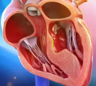
Septal myectomy is a type of open-heart surgery for obstructive hypertrophic cardiomyopathy (HOCM). Hypertrophic cardiomyopathy is a condition where part of the heart muscle (the intraventricular septum) — the wall between the two pumping chambers — becomes abnormally thick. This thickened wall can make the heart’s main exit passage extremely narrow.
When the heart squeezes, the mitral valve may get pulled forward into that narrow passage. As a result, blood has difficulty leaving the heart and getting to the rest of the body, even more so, during exertion. The ‘outflow’ tract through which it leaves the heart and reaches the rest of the body is called the Left Ventricular Outflow Tract (LVOT). A septal myectomy aims to resolve this outflow problem by removing strategically selected septal muscle to widen the outflow tract.
Long-term data or multiple center registries in India are still not comprehensive. However, reports from selected centers seem to match global outcomes. There is a large fall in obstruction and marked symptom improvement. However, Indian centers report higher postoperative gradients compared to global studies. LVOT gradients tell doctors how much extra pressure the left ventricle must generate to push blood through the narrowed outflow tract. Essentially, lower the gradient reading, the better. Global figures show 94% survival at 5 years and 91% at 10 years.
Conditions Treated
Septal myectomy treats conditions that are broadly in the HCOM category. Within this, there are different anatomical forms:
Basal (subaortic) obstructive HOCM: This is the most common form of HCM. The thickening is just below the aortic valve; this narrows the LVOT and also causes systemic anterior motion (SAM) of the mitral valve. This is a condition where the leaflets of the mitral valve are pulled towards the exit (LVOT); as blood rushes through the leaflets may touch the septum and make the exit even narrower.
Diffuse septal hypertrophy: Thickening extends further down the septum, so the surgeon may need a longer, more extensive myectomy.
Mid-ventricular obstructive HCM: Narrowing occurs deeper inside the left ventricle, often near the papillary muscles, rather than directly below the aortic valve.
Apical HCM with a very small LV cavity or apical aneurysm: This is uncommon, but some patients could benefit from transapical (through the apex, or tip of the left ventricle) enlargement or removal of a ventricular obstruction. Apical procedures rely on increasing space inside the ventricle itself, as opposed to the outflow tract. This is relevant when the blockage is too deep or too close to the apex.
HOCM with mitral or papillary muscle abnormalities: When abnormal mitral valve chordae, leaflets or papillary muscles contribute to obstruction, they are corrected in the same surgery.
Indications for Septal Myectomy
The first indications for septal myectomy are symptom based — when a person becomes severely limited by breathlessness, chest discomfort, high levels of fatigue and exercise intolerance. Also, heart failure symptoms like swollen legs or a stomach, and angina. At this stage, basic daily activities like bathing, getting dressed and walking feel like effort.
When the first line of medications don’t usually work, and advanced lines of medication continue to fail, surgical intervention is discussed. In medical terms, the LVOT gradient has to be atleast 50 mmHg. 30 mmHg establishes an ‘obstruction’, but 50 mmHg is the point at which surgery is considered.
When HCM also coexists with another cardiac issue, especially something to do with the mitral valve, surgery becomes a more favoured option, as both issues can be fixed in a single operation. However, these decisions are case-by-case and made in highly experienced HCM centers.
Step-by-Step Guide to Septal Myectomy
- The first step is imaging. Various imaging tests help doctors understand exactly where the heart muscle is thickened, how severe the blockage is, and whether or not the mitral valve and its supporting structures are contributing. TTE (Transthoracic Echocardiocardiogram): This measures septal thickness, LVOT gradient, SAM, and mitral valve leakage, including changes during exercise.
- Cardiac MRI: This gives detailed images of the whole heart muscle, including areas hard to visualize on an echo. It also maps the pattern of thickening and can identify scar tissue and papillary-muscle abnormalities.
- Transesophageal echocardiogram (TEE): This probe-based ultrasound test provides close views of the mitral valve. Cardiac CT: In very select cases, this is used for a very detailed three dimensional view and surgical planning. Once the imaging tests are done, and the operation has been planned out, the patient is placed under general anesthesia. Doctors perform a median sternotomy, hook up the cardiopulmonary bypass machine and arrest the heart. Once the CPB has taken over, the actual operation can begin.
- An aortotomy provides some exposure through the aortic valve into the LVOT. The surgeon then identifies safe ‘landmarks’ near the right coronary cusp and membranous septum. Then, the muscle removal happens — usually a broad, adequately deep segment of basal to mid-septal muscle, extending beyond the mitral-septal contact area.
- After this, the surgeon assesses residual obstruction and SAM, with direct pressure measurements and intraoperative TEE. If necessary, other mitral valve complications may be dealt with. The surgeon may release or remove abnormal supporting ‘strings’ of the mitral valve. They may also loosen, separate and reshape small valve-supporting muscles. Additionally, the mitral valve itself may be repaired if it has any structural problems.
- The aortotomy and sternotomy are closed after de-airing, transferring heart function from the bypass machine and confirming a low residual gradient and acceptable MR.
Advanced Techniques in Septal Myectomy
Extended transaortic myectomy: For more extended blockages. Surgeons remove a thicker section of muscle, often reaching deeper towards the muscles supporting the mitral valve. Mitral valve support repair: As mentioned in the previous section, sometimes the valve may have defects (abnormal strings or muscles), which the surgeon may address in the same procedure.
Mitral valve leaflet repair: If the valve leaflets are a bit longer, or have any other issues, surgeons will address it during the myectomy. While not an innovation within the procedure itself, it extends the scope of issues that septal myectomies can deal with.
Alternate approaches: When the thickened muscle is located very deep in the heart or near the tip (apex), surgeons can enter either through the tip of the heart or mitral valve. This is a bit complex and delicate, often used with great discretion.
Limited-incision surgery: In patients with a favourable anatomy, myectomy can be performed with smaller chest incisions. However, the primary focus is always complete removal and clear access.
Benefits of Septal Myectomy
In highly experienced HCM units, septal myectomy is a highly effective operation. The most immediate benefits revolve around symptom relief. Since the obstruction has been removed, there is increased blood flow to the body — this reduces fatigue, increases exercise tolerance and dramatically reduces breathlessness.
The procedure can make daily life much easier than it used to be. Dizziness and chest discomfort reduce too. The procedure also produces a very sturdy and durable LVOT gradient reduction and commonly reduces SAM-related mitral regurgitation. In India, the average LVOT gradient drops from 113.06 to 15.17 mmHg. Septal thickness reduces from 28.89 to 13.17 mm.
However, it’s crucial to understand the procedure treats the obstruction itself and not the inherited tendency of HCM. For the procedure to have enduring success, ongoing heart-rhythm monitoring, follow up scans and family screening when appropriate. Also, an assessment for ICM (Implantable Cardioverter-Defibrillator) may be recommended.
A myectomy doesn’t eliminate the risk of dangerous heart rhythms, and in select cases, an ICD could be recommended after an individual assessment. Its purpose is to reduce any risk of a sudden cardiac arrest.
Outcomes are generally excellent, with reported clinical success rates above 95% and operative mortality less than 1%.
Risks and Complications
Septal myectomy is a major open-heart surgery. Naturally, it comes with risks and complications. It’s also a very experience-dependent surgery, so specialized HCM centers that perform it routinely are well equipped to deal with any risks.
However, patients and caregivers should understand the potential implications:
- Bleeding or infection: May require a blood transfusion, medicines or occasionally a reoperation.
- Stroke, kidney or lung problems: Standard risks in any major heart surgery, especially in old and frail patients with multiple comorbidities.
- Heart rhythm issues: Some patients develop atrial fibrillation or other rhythm issues.
- Residual blockage or valve leakage: If some narrowing, SAM, or mitral leakage remains, further treatment might be required.
- Low cardiac output: The heart’s ability to pump blood — its ‘capacity’ as a pump — may reduce after the surgery. This can happen even if the surgery itself is successful. In such cases, intensive support may be needed for longer.
- Injury to nearby structures: Extremely rare complications include a hole in the heart’s dividing wall or damage to the aortic valve. Before any surgery, doctors typically engage in a risk stratification process.
- This takes into account ethnicity, family history, comorbidities and multiple other organ parameters to offer a personalized risk level for the procedure. Decisions are always made on the basis of this.
Recovery and Rehab
With cardiac surgeries, rehab is crucial to maximize the benefits. After septal myectomy, the first day or two is usually spent in an ICU (CT-ICU), with teams closely monitoring their blood pressure, cardiac output, kidney function, heart rhythm, breathing and drainage tubes. Most patients are then moved to a regular ward, depending on their recovery. Most patients go home after a week. SAM, LVOT and MR are constantly monitored and patients are cleared for discharge once these have stabilized.
The first few weeks are about cautious living at a gentle pace. Gradual walking is introduced, along with stairs. This helps rebuild aerobic capacity and strength. The wound from a sternotomy takes time to heal, and is still prone to infection, so any activity that may place a stress on the area is off-limits. Heavy lifting, anything requiring ‘max effort’, driving (because of the jerks and seat belt coming over the wound) are to be avoided. Pain medication usually carries on through this period. Breathing exercises become part of daily recovery. Additionally, wound management is crucial.
Bathing itself might be allowed, but in the form of a gentle shower. Doctors have to give a go-ahead for this. Even after they do, scrubbing and vigorous rubbing is not recommended. Unless prescribed, creams, powders and antiseptics are also to be avoided. Bathing in the form of long soaks in a bathtub or hot tub is not recommended. Swimming in pools or the sea isn’t recommended either. Soaking can increase the risk of infection. During this period, red flags include any sort of redness, warmth, swelling, discharge, wound opening, fever or worsening pain.
Once the wound has healed well, a more structured cardiac rehab program can begin. This is usually around the 3 week mark. This would involve progressively increasing exercise loads, introducing very light resistance work and some complex movements, along with heart-rate monitoring when needed and even emotional support. Along the way, a good cardiac rehab program also educates a patient. By the six to eight week mark, life starts feeling ‘normal’ again. Part of this is resuming sexual activity (but with caution about positions and pressure on the wound). Sex can usually be resumed earlier — around the 2-4 week mark — based on how the patient is responding to moderate activity (brisk walking or climbing two flights of stairs) and breathing.
Doctors may then decide how to scale up rehab, or what sort of exercises/sport can be reintroduced, based on the patient’s state. Follow up visits and echocardiograms to check blood-flow pathways, leakage, heart rhythms and function help determine this. Medicines may be adjusted on the basis of the patient's progress.

