

Surgical Heart Defect Closure
 Home
- Surgical Heart Defect Closure


Medically Reviewed By Dr. Vishal Pingle, Cardiothoracic & Heart Transplant Surgeon. Updated on October 19, 2026
Surgical heart defect closure is a broad term for repairs of structural openings or an abnormal blood pathway, mainly caused by a congenital heart defect. Congenital heart defects are structural issues in the heart that are present from birth. The closing itself happens as part of an open-heart surgery, using sutures or a patch. There are a variety of ‘defects’ that surgical closure addresses — atrial septal defects, ventricular septal defects, patent ductus arteriosus, atrioventricular septal defects, partial pulmonary venous connection and other complex defects. In India, surgical guidance recommends early closure for haemodynamically important effects — defects that influence blood flow through the heart. Outcomes tend to vary depending on the patient profile, complexity, comorbidities, heart anatomy, severity, and many other factors. Therefore, it’s hard to offer a broad statistic of success. However, a 2024 study showed mortality rates of 2.87% for VSD (ventricular septal defect) closure.
In general, surgery is preferred when the heart’s anatomy is unsuitable for a catheter device, when multiple defects need correction, or when a durable direct repair is considered safer. According to estimates, based on reports from various pediatric cardiac centers, about 20,000–30,000 congenital heart surgeries are performed annually in India. This estimate includes repairs and operations beyond isolated defect closures. While a reliable number isn’t available, it’s safe to assume the number of isolated defect closure surgeries are lower.
Conditions treated
Not all congenital heart defects require surgery. Small holes may remain harmless or even close on their own. Surgery is recommended when the defect is large, affects heart function and growth, and cannot safely be closed using a catheter-based device.
Atrial septal defects (ASD): A hole in the wall between the heart’s two upper chambers. Extra blood may flow to the lungs and gradually enlarge the right side of the heart.
Ventricular septal defects (VSD): A hole between the two lower pumping chambers. A large VSD can cause fast breathing, frequent chest infections, slow weight gain, poor feeding, or heart failure in infants.
Patent ductus arteriosus: A fetal blood vessel that should close after birth remains open, creating an unwanted connection between the aorta and pulmonary aorta.
Atrioventricular septal defects (AVSD): A large central opening/hole involving both chambers, and often (but not always) abnormal heart valves. This is more common in children with Down’s Syndrome and requires early surgical repair.
Partial anomalous pulmonary venous connection: One or more lung veins drain into the wrong heart chamber or vessel. Surgery reroutes these veins to the left upper chamber (atrium) and often closes an associated ASD.
Complex defects: These are conditions that involve multiple compromised organs. For instance, a condition like tetralogy of Fallot may involve a VSD along with narrowed blood flow to the lungs. In such a situation, surgery would aim to close the hole and improve the route from the heart to the lungs.
There are situations in which surgical closure might actually be counterproductive. In some adults, untreated ASD has allowed high blood pressure to develop in the pulmonary arteries. If this damage becomes severe or permanent, blood can start flowing from the right side of the heart to the left. This reduces oxygen levels.
In such a scenario, the ASD can act as a type of ‘pressure valve’ for the right chamber, which is quite overworked. Closing it could suddenly increase pressure in that chamber and trigger right-sided heart failure.
What the Surgery Involves
A surgical heart defect closure is usually performed under general anesthesia. The patient is fully asleep and feels no pain. Before the procedure, the team reviews a variety of scans — echocardiography, blood work, chest imaging and other tests — to understand the exact location, size and effect of the defect. For the actual procedure itself, the patient is put on a cardiopulmonary bypass machine (CPB). This transfers the function of pumping blood and adding oxygen, temporarily. In the case of an open-heart surgery, an incision is made through the breastbone to reach the heart.
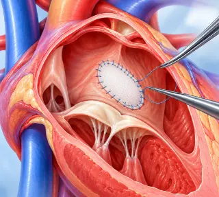
The cardiac surgeon then opens the relevant chamber and closes the abnormal opening. A smaller hole may be closed with surgical stitches, but a very large one may need a patch. The patch is usually made from the patient’s own tissue, animal tissue prepared for medical use or a synthetic patch. The patch becomes part of the heart’s wall over time.
If there are related problems, such as a leaking valve, narrowed blood flow pathway, or abnormally connected lung vein — these can be corrected during the same operation. Where possible, heart surgeons like to resolve multiple issues during a single operation, as repeated surgeries can be very stressful for the body. Before completing the operation, the team checks the repair with ultrasound imaging (transesophageal echocardiography) of the heart. They look for remaining leaks, valve issues or any blood flow problems.
The heart is then restarted as the CPB is slowly weaned off. Finally, the breastbone and skin are closed. The patient is then transferred to a cardiac ICU for close monitoring of heart rhythm, breathing, blood pressure, pain, fluid balance and bleeding. The duration of ICU stay will depend on the specifics of the case — the type of defect, the patient’s age, and whether the repair is simple or complex.
In some ‘straightforward’ cases involving an ASD, some VSDs, partial anomalous venous connection or select atrioventricular septal defects, a minimally invasive approach may be considered. But this approach is not suitable for all: very small patients, complex anatomy, severe pulmonary hypertension or a need for multiple repairs may make an open surgery safer.
Benefits and Trade offs
In the most severe cases, a surgical heart defect closure can save a life. It can relieve breathlessness, reduce abnormal blood flow, recurrent chest infections, and help prevent progressive chamber enlargement, arrhythmias, pulmonary hypertension, valve injury or infective endocarditis. Essentially, a barrage of heart complications. However, there are tradeoffs. These surgeries come with plenty of potential complications, especially in very young patients. Common early issues include bleeding that may require a blood transfusion, temporary breathing support, fluid buildup around the lungs or heart, and wound and chest infection. Some patients develop an abnormal heart rhythm — in most cases, this settles with medication, but a small number might need a pacemaker.
There are more uncommon but serious complications such as a residual shunt (a small remaining hole) stroke, reduced cardiac function, blood clots, leakage of lymph fluid into the chest (chylothorax), or kidney issues. Death is rare in straightforward cases, but that risk substantially rises with complex defects. Global outcomes show a 0.3% operative mortality rate for patch closure of ASD and 0.9% for patch closure of VSD. Indian studies report an estimate of 5.63% across different congenital heart operations. Risk and outcomes vary dramatically, based on the operation type and patient profile.
Recovery and Rehab
Recovery begins in the ICU with teams checking vitals, especially bleeding, fluid balance, heart rhythm, blood pressure, bleeding and pain. In straightforward cases, patients move to a ward after a couple of days, but the timeline is longer for newborns, complex defects or any patient with complications.
Early rehab focuses on sitting up, deep-breathing exercises, walking, pain control, wound care and good nutrition. Children may require feeding support, and in some cases speech therapy, extended physio, or developmental follow ups.
Until the surgeon confirms that the breastbone has healed, heavy lifting, pushing and pulling, and contact sports are to be avoided. Normally, this is around 6-8 weeks. Gentle walking with gradual progressions and normal daily activity is encouraged. Follow up visits are extremely crucial. An ECG and echocardiogram to check heart rhythm, healing and the success of the repair.
Cardiac rehab is part of the process, but not in the more standardized way that follows other procedures. For adults, there are some structured aspects, but the overall approach is highly personalized based on the patient’s profile and multiple issues. For children, however, rehab is broader and far more play-based. For kids with prolonged illnesses, occupational, speech and developmental therapy are crucial. Emotional support for return to nursery, school, sport and confidence with peer groups is crucial and integrated within the larger rehab focus. Supervised pediatric programmes may combine breathwork, muscle strengthening, aerobic activity and cool-downs as part of a session, to be done 2-3 times a week, for a window of 12 weeks.
Children with a successful repair of a simple defect gradually return to normal play. Competitive sport may, however, require an echocardiogram, ECG and sometimes, an exercise test. The studies are very encouraging — one study of children with congenital heart defects showed that 57% took up competitive sport, and 72% played some sport recreationally. For children with simple ASD or VSD closures with no residual complications, guidelines generally allow a return to all sports within 3-6 months.
Even children with developmental delays often improve with the right support.

