CaseStudy

TMVR HVSGhatkopar

Hybrid Approach - TMVR & Angioplasty

An 80-year-old male patient presented to HVS Superspeciality Hospital with acute severe mitral regurgitation (MR) and complex coronary artery disease (CAD). The case was high-risk, involving multiple critical cardiac pathologies:


  • Mitral Valve Disease: Acute, severe MR
  • Coronary Artery Disease:
    • Left Anterior Descending (LAD): Total occlusion, deemed non-viable
    • Large Obtuse Marginal (OM) and Left Circumflex (LCX) arteries: Significant disease


The clinical challenge lay in managing both the valvular and ischemic burden, especially considering the patient's advanced age and comorbid status.


Treatment Plan:

1. Percutaneous Coronary Intervention (PCI) to LCX and OM — aimed at relieving the ischemic burden.

2. Staged Mitral Valve Repair using MitraClip therapy (Indian brand: Meril’s MyClip) — a revolutionary transcatheter edge-to-edge repair (TEER) system designed to reduce MR without the need for open-heart surgery.


Procedure 1: Angioplasty of LCX and OM

The first stage involved successful angioplasty and stenting of the LCX and OM arteries. The LAD was found to be non-viable, and hence not revascularized. The intervention restored perfusion to large myocardial territories and stabilized the patient for the second stage.

Outcome: Procedure was uneventful, and the patient showed significant symptomatic relief from angina and dyspnea.


Procedure 2: Mitral Valve Repair with MyClip

After cardiac stabilization, a transcatheter mitral valve repair was performed using the MyClip system by Meril Life Sciences — a cutting-edge Indian technology allowing precise, edge-to-edge leaflet approximation.

  • A long and wide clip was chosen to effectively cover the regurgitant jet.
  • Transesophageal echocardiography (TEE) guidance was used throughout the procedure.


Result: Post-clip deployment, there was only trivial residual MR, and the patient showed dramatic clinical improvement.


Team of Experts

This complex, staged hybrid intervention was made possible by the synergy of an expert multi-disciplinary team:

Interventional Cardiologists

  • Dr. Meghav Shah
  • Dr. Ankur Phatarpekar
  • Dr. Amit Gangawani
  • Dr. Harshad Sagar
  • Dr. Kunal Patankar


Interventional Imaging Specialists

  • Dr. Gourish Shinde
  • Dr. Pravin Lovhale


Conclusion

This case highlights the power of team-based, staged, and minimally invasive therapy for managing complex cardiac disease in elderly patients. By combining revascularization with transcatheter mitral repair, the heart team at Heart & Vascular Superspeciality Hospitals was able to provide a new lease of life to a high-risk, elderly patient — without open-heart surgery.

Tavip Casestudy

Tavi Procedure

A 67 years old male patient with a history of dyspnoea of exertion since 1 month which worsened since 15 days. He was diagnosed with severe Aortic Valve Stenosis.

On Examination

Vitals Stable

CVS – EFM Systolic Murmur

Management/ Progress in Hospital

He underwent TAVI (Trancatheter Aortic Valve Implantation) under local Anaesthisia + Sedation by Dr. Ankur Phatarpekar and his team through right femorial artery.

Post procedure was shifted to ICU with stable Haemodynamic for observation.

Post procedure ECG sinus rhythm no ST-T changes.

After 24hrs left femorial sheaths were removed.

He was mobilised out of the bed.

Beta Blockers with held in view of Borderline Bradycardia.

Post Procedure Echo Screening.

Investigation

Pre-Operative

2D-Echo – Gross Concentric LVH. All chambers normal size. Mild and distal septum mildly hypokinetic. Heavily calcified Aortic valve with reduced opening with peak by mean gradient of 86/53 mm of Hg suggestive of Severe AS with Grade ¼ AR. Grade ¼ MR, Grade ¼ AR. No evidence of PR/TR. No significant gradient across LVOT/RVOT. Normal LVEF – 61%

Post- Operative

2D-ECHO- Normal LV systolic function. k/c/o degenerative Severe AS , s/p TAVI with normally functioning of Aortic valve. Mild Concentric LVH. All chambers normal size.

Bioprosthetic Aortic valve in situ, opening well. No e/o paravalvular leak.

Grade I MR. No evidence of AR/PR/TR. Peak by mean gradient across Aortic valve is 28/16 mm of Hg. AVA by continuity is 1.43 sq.cm.

LVEF 65%.

Operative Notes

Right femorial and left femorial artery venous punctured.

Left femorial side 6-F sheath introduced in both venous and artery.

Right common femorial artery 10-F sheath introduced.

23 size balloon used for commissurotomy and inflated for 3 seconds.

Ventricle with pacing at 180 beats/min gradiant aortic valve – nil.

Following commissurotomy 23 size MyVal pericardial, bioprothes crimpel introduced and implanted across aortic valve with ventricular pacing of 180 beats/min.

Result – Valve position intra-annular

No para valvular leak.

Good result.

Right femorial cannulation closed with proglide.

2 proglide used.

Good haemostasis.

Patient tolerated procedure well.

Case Study 2

Glue Embolization of Lower Limb

Procedure/Surgery Name: Endovascular Glue Embolization of lower limb/leg Arterio-Venous Malformation (AVM)

Doctors: Dr. Bhavesh Arun Popat, Dr. Karan M Anandpara

Pre-Op Details & Diagnosis

A 29 year old female

  • Progressively increasing painful swelling on anterior aspect of right distal leg of 4 months duration
  • No skin changes
  • Minimal right distal leg edema
  • Tender on palpation +
  • Distal pulses (ATA, DPA and PTA) palpable +

Investigation

  • Local Ultrasound and Doppler – echogenic ill defined soft tissue area with few serpiginous vascular channels within s/o vascular malformation
  • MRI – confirmed the findings of USG; arterial feeders with intralesional nidus noted s/o likely arterio-venous malformation (AVM)

Angiography Findings:

  • Abnormal leash of vessels
  • Internal nidus
  • Atleast 4 arterial feeders from anterior tibial artery
  • Early draining vein – washout from accompanying anterior tibial veins
  • Distal main anterior tibial artery and DPA shows slow, but antegrade adequate distal flow

Diagnosis

Complex arterio-veneous malformation (avm) of lower limb/leg with multiple arterial feeders from anterior tibial artery.

Treatment Plan

  • Staged; combined ultrasound and fluoroscopic percutaneous and sos endovascular glue embolisation
  • Followed by surgical excision

Procedure Details

  • Percutaneous USG guided access taken
  • Catheter in the right popliteal artery via left CFA access
  • Endovascular contrast injection to localize the nidus and to monitor residual lesion
  • Following this, multiple percutaneous access taken to target various components of the AVM
  • Manual hand compression given to prevent inadvertent reflux into the draining vein
  • Endovascular embolization attempted, but ultimately was not done due to high probability of reflux into the main anterior tibial artery due to intra-nidus vascular communication. Therefore, percutaneous embolization done with Glue

Post Procedure DSA

  • Near complete exclusion of the AVM
  • Glue cast visible

Conclusion

  • Essential to classify vascular malformations appropriately – Venous, lymphatic, AVM, combined vascular malformations
  • Set ‘realistic expectations’
  • Multiple procedures
  • Difficult to treat completely
  • Team Work
Case Study

Geniculate Artery Embolization

Procedure/Surgery Name: Geniculate Artery embolization for Recurrent Hemarthrosis

Doctors: Dr Bhavesh Arun Popat, Dr Karan M Anandpara

Pre-Op Details & Diagnosis

A 72 years old male

Comorbidities: Morbidly obese, poorly controlled diabetes, hypertensive, previous H/O CABG.

Complaints: Bilateral knee pain, joint aches and soreness (left>right knee).

Pain after joint overuse.

Reduced physical activity and quality of life.

Local knee tenderness +.

X ray bilateral knee s/o bilateral osteoarthritic changes (left more than right).

H/O left sided recurrent hemarthrosis for which percutaneous USG guided aspiration of fluid done outside.

USG local part suggestive of left sided hemarthrosis – hyperechoeic fluid with septations in the left knee joint cavity.

MRI done s/o changes of severe osteoarthritis with left sided hemarthrosis

Pain not relieved by conservative medical management i.e. by anti-inflammatory drugs or corticosteroids.

Not relieved by intra-articular steroid injection.

Not a candidate for joint replacement/arthroplasty in view of significant medical comorbidities, contraindications for general anaesthesia and patient’s refusal for knee arthroplasty.

Treatment Plan

Planned for Left sided Geniculate Artery Embolization in view of recurrent hemarthrosis and left joint pain.

Details about the Procedure/Surgery:

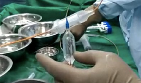
Procedure under local anesthesia. Groin prepared.

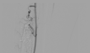
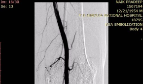
Left Common Femoral Artery antegrade access.

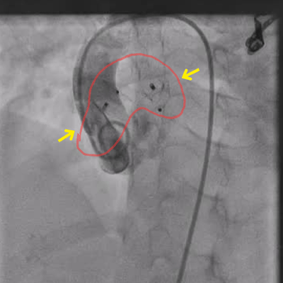
DSA Angiogram done which showed hypertrophied superior and inferior medial geniculate arteries.

Synovial vascular blush noted.

Super selective cannulation of the superior and inferior geniculate arteries done using Progreat microcatheter.

Embolization done with PVA particles.

Post Procedure Details:

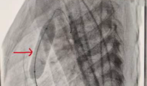
Post procedure DSA showed reduction in the synovial vascularity. Reduced arterial blush noted.

Sheath removed by manual compression.

Patient discharged on same day.

Significant reduction of pain on 1 and 3 month follow up.

Conclusion

Geniculate artery embolization (GAE) reduces pain of osteoarthritis by decreasing the vascular supply to the hypertrophied and inflamed synovium and has a role in cases of recurrent hemarthrosis.

It is safe, effective and minimally invasive.

No cuts, no stitches are required.

It can be Performed under local anesthesia.

No general anaesthesia needed.

It is a day care procedure with a same day discharge.